38
views
0
recommends
+1 Recommend
0 collections
    0
    shares
      • Record: found
      • Abstract: found
      • Article: found
      Is Open Access

      Quantitative properties and receptor reserve of the DAG and PKC branch of G q-coupled receptor signaling

      research-article

      Read this article at

      Bookmark
          There is no author summary for this article yet. Authors can add summaries to their articles on ScienceOpen to make them more accessible to a non-specialist audience.

          Abstract

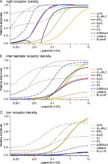
          G q protein–coupled receptors (G qPCRs) of the plasma membrane activate the phospholipase C (PLC) signaling cascade. PLC cleaves the membrane lipid phosphatidylinositol 4,5-bisphosphate (PIP 2) into the second messengers diacylgycerol (DAG) and inositol 1,4,5-trisphosphate (IP 3), leading to calcium release, protein kinase C (PKC) activation, and in some cases, PIP 2 depletion. We determine the kinetics of each of these downstream endpoints and also ask which is responsible for the inhibition of KCNQ2/3 (K V7.2/7.3) potassium channels in single living tsA-201 cells. We measure DAG production and PKC activity by Förster resonance energy transfer–based sensors, and PIP 2 by KCNQ2/3 channels. Fully activating endogenous purinergic receptors by uridine 5′triphosphate (UTP) leads to calcium release, DAG production, and PKC activation, but no net PIP 2 depletion. Fully activating high-density transfected muscarinic receptors (M 1Rs) by oxotremorine-M (Oxo-M) leads to similar calcium, DAG, and PKC signals, but PIP 2 is depleted. KCNQ2/3 channels are inhibited by the Oxo-M treatment (85%) and not by UTP (<1%), indicating that depletion of PIP 2 is required to inhibit KCNQ2/3 in response to receptor activation. Overexpression of A kinase–anchoring protein (AKAP)79 or calmodulin (CaM) does not increase KCNQ2/3 inhibition by UTP. From these results and measurements of IP 3 and calcium presented in our companion paper (Dickson et al. 2013. J. Gen. Physiol. http://dx.doi.org/10.1085/jgp.201210886), we extend our kinetic model for signaling from M 1Rs to DAG/PKC and IP 3/calcium signaling. We conclude that calcium/CaM and PKC-mediated phosphorylation do not underlie dynamic KCNQ2/3 channel inhibition during G qPCR activation in tsA-201 cells. Finally, our experimental data provide indirect evidence for cleavage of PI(4)P by PLC in living cells, and our modeling revisits/explains the concept of receptor reserve with measurements from all steps of G qPCR signaling.

          Related collections

          Most cited references58

          • Record: found
          • Abstract: found
          • Article: not found

          Inositol trisphosphate receptor Ca2+ release channels.

          The inositol 1,4,5-trisphosphate (InsP3) receptors (InsP3Rs) are a family of Ca2+ release channels localized predominately in the endoplasmic reticulum of all cell types. They function to release Ca2+ into the cytoplasm in response to InsP3 produced by diverse stimuli, generating complex local and global Ca2+ signals that regulate numerous cell physiological processes ranging from gene transcription to secretion to learning and memory. The InsP3R is a calcium-selective cation channel whose gating is regulated not only by InsP3, but by other ligands as well, in particular cytoplasmic Ca2+. Over the last decade, detailed quantitative studies of InsP3R channel function and its regulation by ligands and interacting proteins have provided new insights into a remarkable richness of channel regulation and of the structural aspects that underlie signal transduction and permeation. Here, we focus on these developments and review and synthesize the literature regarding the structure and single-channel properties of the InsP3R.
            Bookmark
            • Record: found
            • Abstract: found
            • Article: not found

            Rapid chemically induced changes of PtdIns(4,5)P2 gate KCNQ ion channels.

            To resolve the controversy about messengers regulating KCNQ ion channels during phospholipase C-mediated suppression of current, we designed translocatable enzymes that quickly alter the phosphoinositide composition of the plasma membrane after application of a chemical cue. The KCNQ current falls rapidly to zero when phosphatidylinositol 4,5-bisphosphate [PtdIns(4,5)P2 or PI(4,5)P2] is depleted without changing Ca2+, diacylglycerol, or inositol 1,4,5-trisphosphate. Current rises by 30% when PI(4,5)P2 is overproduced and does not change when phosphatidylinositol 3,4,5-trisphosphate is raised. Hence, the depletion of PI(4,5)P2 suffices to suppress current fully, and other second messengers are not needed. Our approach is ideally suited to study biological signaling networks involving membrane phosphoinositides.
              Bookmark
              • Record: found
              • Abstract: found
              • Article: not found

              A genetically encoded fluorescent reporter reveals oscillatory phosphorylation by protein kinase C

              Signals transduced by kinases depend on the extent and duration of substrate phosphorylation. We generated genetically encoded fluorescent reporters for PKC activity that reversibly respond to stimuli activating PKC. Specifically, phosphorylation of the reporter expressed in mammalian cells causes changes in fluorescence resonance energy transfer (FRET), allowing real time imaging of phosphorylation resulting from PKC activation. Targeting of the reporter to the plasma membrane, where PKC is activated, reveals oscillatory phosphorylation in HeLa cells in response to histamine. Each oscillation in substrate phosphorylation follows a calcium oscillation with a lag of ∼10 s. Novel FRET-based reporters for PKC translocation, phosphoinositide bisphosphate conversion to IP3, and diacylglycerol show that in HeLa cells the oscillatory phosphorylations correlate with Ca2+-controlled translocation of conventional PKC to the membrane without oscillations of PLC activity or diacylglycerol. However, in MDCK cells stimulated with ATP, PLC and diacylglycerol fluctuate together with Ca2+ and phosphorylation. Thus, specificity of PKC signaling depends on the local second messenger-controlled equilibrium between kinase and phosphatase activities to result in strict calcium-controlled temporal regulation of substrate phosphorylation.
                Bookmark

                Author and article information

                Journal
                J Gen Physiol
                J. Gen. Physiol
                jgp
                The Journal of General Physiology
                The Rockefeller University Press
                0022-1295
                1540-7748
                May 2013
                : 141
                : 5
                : 537-555
                Affiliations
                [1 ]Department of Physiology and Biophysics, University of Washington, Seattle, WA 98195
                [2 ]Department of Neurology, RWTH Aachen University, 52062 Aachen, Germany
                Author notes
                Correspondence to Bertil Hille: hille@ 123456u.washington.edu

                B.H. Falkenburger and E.J. Dickson contributed equally to this paper.

                Article
                201210887
                10.1085/jgp.201210887
                3639584
                23630338
                3cc1007c-719a-4a0a-bc0d-48561eb47d44
                © 2013 Falkenburger et al.

                This article is distributed under the terms of an Attribution–Noncommercial–Share Alike–No Mirror Sites license for the first six months after the publication date (see http://www.rupress.org/terms). After six months it is available under a Creative Commons License (Attribution–Noncommercial–Share Alike 3.0 Unported license, as described at http://creativecommons.org/licenses/by-nc-sa/3.0/).

                History
                : 23 August 2012
                : 26 March 2013
                Categories
                Research Article

                Anatomy & Physiology
                Anatomy & Physiology

                Comments

                Comment on this article