14
views
0
recommends
+1 Recommend
0 collections
    0
    shares
      • Record: found
      • Abstract: found
      • Article: found
      Is Open Access

      Establishment of reference values of the caudal vena cava by fast‐ultrasonography through different views in healthy dogs

      research-article

      Read this article at

      Bookmark
          There is no author summary for this article yet. Authors can add summaries to their articles on ScienceOpen to make them more accessible to a non-specialist audience.

          Abstract

          Background

          Clinical assessment of intravascular volume status is challenging. In humans, ultrasonographic assessment of the inferior vena cava diameter, directly or as a ratio to the aortic diameter is used to estimate intravascular volume status.

          Objectives

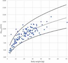
          To ultrasonographically obtain reference values (RV) for caudal vena cava diameter (CVC D), area (CVC a) and aortic ratios using 3 views in awake healthy dogs.

          Animals

          One hundred and twenty‐six healthy adult dogs from clients, students, faculty, or staff.

          Methods

          Prospective, multicenter, observational study. Two observer pairs evaluated CVC D by a longitudinal subxiphoid view (SV), a transverse 11th‐13th right hepatic intercostal view (HV), and a longitudinal right paralumbar view (PV). Inter‐rater agreements were estimated using concordance correlation coefficients (CCC). For body weight (BW) dependent variables, RVs were calculated using allometric scaling for variables with a CCC ≥ 0.7.

          Results

          The CCC was ≤0.43 for the CVC/aorta ratio at the PV and ≤0.43 in both inspiration and expiration for CVC at the SV. The RVs using allometric scaling for CVC a at the HV for inspiration, expiration, and for CVC D at the PV were 6.16 × BW 0.762, 7.24 × BW 0.787, 2.79 × BW 0.390, respectively.

          Conclusions and Clinical Importance

          The CVC D, measured at the HV and PV in healthy awake dogs of various breeds has good inter‐rater agreement suggesting these sites are reliable in measuring CVC D. Established RVs for CVC D for these sites need further comparison to results obtained in hypovolemic and hypervolemic dogs to determine their usefulness to evaluate volume status in dogs.

          Related collections

          Most cited references36

          • Record: found
          • Abstract: found
          • Article: not found

          A concordance correlation coefficient to evaluate reproducibility.

          L Lin (1989)
          A new reproducibility index is developed and studied. This index is the correlation between the two readings that fall on the 45 degree line through the origin. It is simple to use and possesses desirable properties. The statistical properties of this estimate can be satisfactorily evaluated using an inverse hyperbolic tangent transformation. A Monte Carlo experiment with 5,000 runs was performed to confirm the estimate's validity. An application using actual data is given.
            Bookmark
            • Record: found
            • Abstract: found
            • Article: not found

            Does central venous pressure predict fluid responsiveness? A systematic review of the literature and the tale of seven mares.

            Central venous pressure (CVP) is used almost universally to guide fluid therapy in hospitalized patients. Both historical and recent data suggest that this approach may be flawed. A systematic review of the literature to determine the following: (1) the relationship between CVP and blood volume, (2) the ability of CVP to predict fluid responsiveness, and (3) the ability of the change in CVP (DeltaCVP) to predict fluid responsiveness. MEDLINE, Embase, Cochrane Register of Controlled Trials, and citation review of relevant primary and review articles. Reported clinical trials that evaluated either the relationship between CVP and blood volume or reported the associated between CVP/DeltaCVP and the change in stroke volume/cardiac index following a fluid challenge. From 213 articles screened, 24 studies met our inclusion criteria and were included for data extraction. The studies included human adult subjects, healthy control subjects, and ICU and operating room patients. Data were abstracted on study design, study size, study setting, patient population, correlation coefficient between CVP and blood volume, correlation coefficient (or receive operator characteristic [ROC]) between CVP/DeltaCVP and change in stroke index/cardiac index, percentage of patients who responded to a fluid challenge, and baseline CVP of the fluid responders and nonresponders. Metaanalytic techniques were used to pool data. The 24 studies included 803 patients; 5 studies compared CVP with measured circulating blood volume, while 19 studies determined the relationship between CVP/DeltaCVP and change in cardiac performance following a fluid challenge. The pooled correlation coefficient between CVP and measured blood volume was 0.16 (95% confidence interval [CI], 0.03 to 0.28). Overall, 56+/-16% of the patients included in this review responded to a fluid challenge. The pooled correlation coefficient between baseline CVP and change in stroke index/cardiac index was 0.18 (95% CI, 0.08 to 0.28). The pooled area under the ROC curve was 0.56 (95% CI, 0.51 to 0.61). The pooled correlation between DeltaCVP and change in stroke index/cardiac index was 0.11 (95% CI, 0.015 to 0.21). Baseline CVP was 8.7+/-2.32 mm Hg [mean+/-SD] in the responders as compared to 9.7+/-2.2 mm Hg in nonresponders (not significant). This systematic review demonstrated a very poor relationship between CVP and blood volume as well as the inability of CVP/DeltaCVP to predict the hemodynamic response to a fluid challenge. CVP should not be used to make clinical decisions regarding fluid management.
              Bookmark
              • Record: found
              • Abstract: found
              • Article: not found

              Intensivist use of hand-carried ultrasonography to measure IVC collapsibility in estimating intravascular volume status: correlations with CVP.

              Volume status assessment is an important aspect of patient management in the surgical intensive care unit (SICU). Echocardiologist-performed measurement of IVC collapsibility index (IVC-CI) provides useful information about filling pressures, but is limited by its portability, cost, and availability. Intensivist-performed bedside ultrasonography (INBU) examinations have the potential to overcome these impediments. We used INBU to evaluate hemodynamic status of SICU patients, focusing on correlations between IVC-CI and CVP. Prospective evaluation of hemodynamic status was conducted on a convenience sample of SICU patients with a brief (3 to 10 minutes) INBU examination. INBU examinations were performed by noncardiologists after 3 hours of didactics in interpreting and acquiring two-dimensional and M-mode images, and > or =25 proctored examinations. IVC-CI measurements were compared with invasive CVP values. Of 124 enrolled patients, 101 had CVP catheters (55 men, mean age 58.3 years, 44.6% intubated). Of these, 18 patients had uninterpretable INBU examinations, leaving 83 patients with both CVP monitoring devices and INBU IVC evaluations. Patients in three IVC-CI ranges ( 0.60) demonstrated significant decrease in mean CVP as IVC-CI increased (p = 0.023). Although 40% of this group had a CVP >12 mmHg. Conversely, >60% of patients with IVC-CI >0.6 had CVP 0.60) collapsibility ranges. Additional studies are needed to confirm and expand on findings of this study.
                Bookmark

                Author and article information

                Contributors
                kris.gommeren@uliege.be
                Journal
                J Vet Intern Med
                J. Vet. Intern. Med
                10.1111/(ISSN)1939-1676
                JVIM
                Journal of Veterinary Internal Medicine
                John Wiley and Sons Inc. (Hoboken )
                0891-6640
                1939-1676
                10 May 2018
                Jul-Aug 2018
                : 32
                : 4 ( doiID: 10.1111/jvim.2018.32.issue-4 )
                : 1308-1318
                Affiliations
                [ 1 ] Department of Clinical Sciences Faculty of Veterinary Medicine, University of Liège Belgium
                [ 2 ] Department of Veterinary Clinical and Diagnotic Sciences Faculty of Veterinary Medicine, University of Calgary Canada
                [ 3 ] U955‐IMRB, INSERM, Ecole Nationale Vétérinaire d'Alfort, UPEC Maisons‐Alfort France
                Author notes
                [*] [* ] Correspondence Kris Gommeren, CVU Liège, Quartier Vallée 2, Avenue de Cureghem, Sart Tilman 4000 Liège, Belgium. Email: kris.gommeren@ 123456uliege.be
                Author information
                http://orcid.org/0000-0002-3433-0767
                http://orcid.org/0000-0002-0936-4834
                Article
                JVIM15136
                10.1111/jvim.15136
                6060313
                29749656
                400e6150-803e-4bfe-a020-ada916e61c98
                Copyright © 2018 The Authors. Journal of Veterinary Internal Medicine published by Wiley Periodicals, Inc. on behalf of the American College of Veterinary Internal Medicine.

                This is an open access article under the terms of the http://creativecommons.org/licenses/by-nc/4.0/ License, which permits use, distribution and reproduction in any medium, provided the original work is properly cited and is not used for commercial purposes.

                History
                : 21 December 2017
                : 14 February 2018
                : 27 March 2018
                Page count
                Figures: 15, Tables: 4, Pages: 11, Words: 6906
                Funding
                Funded by: Scil Animal Care Company
                Categories
                Standard Article
                SMALL ANIMAL
                Standard Articles
                Cardiology
                Custom metadata
                2.0
                jvim15136
                July/August 2018
                Converter:WILEY_ML3GV2_TO_NLMPMC version:version=5.4.4 mode:remove_FC converted:26.07.2018

                Veterinary medicine
                allometric scaling,caudal vena cava,fast echography,intravascular volume status

                Comments

                Comment on this article