22
views
0
recommends
+1 Recommend
0 collections
    0
    shares
      • Record: found
      • Abstract: found
      • Article: found
      Is Open Access

      Eldecalcitol, an Active Vitamin D 3 Derivative, Prevents Trabecular Bone Loss and Bone Fragility in Type I Diabetic Model Rats

      research-article

      Read this article at

      Bookmark
          There is no author summary for this article yet. Authors can add summaries to their articles on ScienceOpen to make them more accessible to a non-specialist audience.

          Abstract

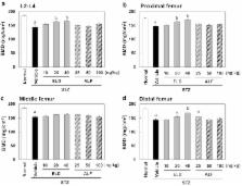
          Diabetes mellitus is known to adversely affect the bones and be associated with increased fracture risk. We examined whether eldecalcitol (ELD), an active vitamin D 3 derivative, could inhibit the diabetic bone loss in streptozotocin-induced type I diabetic rats. ELD (10, 20, or 40 ng/kg), alfacalcidol (ALF; 25, 50, or 100 ng/kg), or vehicle was administered 5 times per week for 12 weeks from 1 week after diabetes induction. Normal control rats received the vehicle. Bone turnover markers, bone mineral density (BMD), and biomechanical strength of the lumbar spine and femur were measured, and bone histomorphometry was performed. Content of advanced glycation end products (AGEs) in the femoral shaft was also determined. In diabetic rats, serum osteocalcin (OC) concentration was lower and urinary excretion of deoxypyridinoline (DPD) tended to be higher than in normal rats. Areal BMD and maximum load of the lumbar vertebrae and femoral shaft were lower in diabetic rats than in normal rats. All doses of ELD and the highest dose of ALF reduced urinary DPD excretion, but had no effect on serum OC. The 20 and 40 ng/kg doses of ELD prevented decreases in BMD and the highest dose of ELD prevented the reduction in maximum load of the lumbar vertebrae, while ALF did not change these parameters. ELD and ALF did not affect areal BMD or biomechanical strength of the femoral shaft. In diabetic rats, bone volume and trabecular thickness in the trabecular bone of the lumbar vertebrae decreased and trabecular separation increased compared to normal rats. ELD and ALF prevented diabetes-induced deterioration of trabecular microstructure. AGE content in the femoral cortical bone increased in the diabetic rats, and ELD and ALF did not change AGE content compared to the diabetic rats. These results indicated that ELD suppressed bone resorption and prevented trabecular bone loss and deterioration of trabecular microstructure, resulting in prevention of reduction in biomechanical strength in type I diabetic rats.

          Related collections

          Most cited references33

          • Record: found
          • Abstract: found
          • Article: not found

          Collagen cross-links as a determinant of bone quality: a possible explanation for bone fragility in aging, osteoporosis, and diabetes mellitus.

          Collagen cross-linking, a major post-translational modification of collagen, plays important roles in the biological and biomechanical features of bone. Collagen cross-links can be divided into lysyl hydroxylase and lysyloxidase-mediated enzymatic immature divalent cross-links,mature trivalent pyridinoline and pyrrole cross-links, and glycation- or oxidation-induced non-enzymatic cross-links(advanced glycation end products) such as glucosepane and pentosidine. These types of cross-links differ in the mechanism of formation and in function. Material properties of newly synthesized collagen matrix may differ in tissue maturity and senescence from older matrix in terms of crosslink formation. Additionally, newly synthesized matrix in osteoporotic patients or diabetic patients may not necessarily be as well-made as age-matched healthy subjects. Data have accumulated that collagen cross-link formation affects not only the mineralization process but also microdamage formation. Consequently, collagen cross-linking is thought to affect the mechanical properties of bone. Furthermore,recent basic and clinical investigations of collagen cross-links seem to face a new era. For instance, serum or urine pentosidine levels are now being used to estimate future fracture risk in osteoporosis and diabetes. In this review, we describe age-related changes in collagen cross-links in bone and abnormalities of cross-links in osteoporosis and diabetes that have been reported in the literature.
            Bookmark
            • Record: found
            • Abstract: found
            • Article: not found

            Diabetic neuropathy--a review.

            Diabetic neuropathy is the most common neuropathy in industrialized countries, and it is associated with a wide range of clinical manifestations. The vast majority of patients with clinical diabetic neuropathy have a distal symmetrical form of the disorder that progresses following a fiber-length-dependent pattern, with sensory and autonomic manifestations predominating. This pattern of neuropathy is associated with a progressive distal axonopathy. Patients experience pain, trophic changes in the feet, and autonomic disturbances. Occasionally, patients with diabetes can develop focal and multifocal neuropathies that include cranial nerve involvement and limb and truncal neuropathies. This neuropathic pattern tends to occur after 50 years of age, and mostly in patients with long-standing diabetes mellitus. Length-dependent diabetic polyneuropathy does not show any trend towards improvement, and either relentlessly progresses or remains relatively stable over a number of years. Conversely, the focal diabetic neuropathies, which are often associated with inflammatory vasculopathy on nerve biopsies, remain self-limited, sometimes after a relapsing course.
              Bookmark
              • Record: found
              • Abstract: found
              • Article: not found

              Osteoporosis in patients with diabetes mellitus.

              Demographic trends with longer life expectancy and a lifestyle characterized by low physical activity and high-energy food intake contribute to an increasing incidence of diabetes mellitus and osteoporosis. Diabetes mellitus is a risk factor for osteoporotic fractures. Patients with recent onset of type 1 diabetes mellitus may have impaired bone formation because of the absence of the anabolic effects of insulin and amylin, whereas in long-standing type 1 diabetes mellitus, vascular complications may account for low bone mass and increased fracture risk. Patients with type 2 diabetes mellitus display an increased fracture risk despite a higher BMD, which is mainly attributable to the increased risk of falling. Strategies to improve BMD and to prevent osteoporotic fractures in patients with type 1 diabetes mellitus may include optimal glycemic control and aggressive prevention and treatment of vascular complications. Patients with type 2 diabetes mellitus may additionally benefit from early visual assessment, regular exercise to improve muscle strength and balance, and specific measures for preventing falls.
                Bookmark

                Author and article information

                Contributors
                +81-3-3273-2613 , endokui@chugai-pharm.co.jp
                Journal
                Calcif Tissue Int
                Calcif. Tissue Int
                Calcified Tissue International
                Springer US (New York )
                0171-967X
                1432-0827
                17 June 2017
                17 June 2017
                2017
                : 101
                : 4
                : 433-444
                Affiliations
                [1 ]GRID grid.418587.7, Product Research Department, Fuji Gotemba Research Laboratories, , Chugai Pharmaceutical Co., Ltd, ; 1-135 Komakado, Gotemba, Shizuoka, 412-8513 Japan
                [2 ]ISNI 0000 0001 0661 2073, GRID grid.411898.d, Department of Orthopedic Surgery, , Jikei University School of Medicine, ; 3-25-8 Nishishinbashi, Minato-ku, Tokyo, 105-8461 Japan
                [3 ]GRID grid.418587.7, Medical Science Department, , Chugai Pharmaceutical Co., Ltd, ; 2-1-1 Nihombashi Muromachi, Chuo-ku, Tokyo, 103-8324 Japan
                Article
                298
                10.1007/s00223-017-0298-8
                5587631
                28624935
                43db3a83-7dcc-45de-9a2c-83fd14ad345c
                © The Author(s) 2017

                Open AccessThis article is distributed under the terms of the Creative Commons Attribution 4.0 International License ( http://creativecommons.org/licenses/by/4.0/), which permits unrestricted use, distribution, and reproduction in any medium, provided you give appropriate credit to the original author(s) and the source, provide a link to the Creative Commons license, and indicate if changes were made.

                History
                : 7 February 2017
                : 7 June 2017
                Categories
                Original Research
                Custom metadata
                © Springer Science+Business Media, LLC 2017

                Human biology
                eldecalcitol,diabetic osteoporosis,streptozotocin,bone quality
                Human biology
                eldecalcitol, diabetic osteoporosis, streptozotocin, bone quality

                Comments

                Comment on this article