31
views
0
recommends
+1 Recommend
0 collections
    0
    shares
      • Record: found
      • Abstract: found
      • Article: found
      Is Open Access

      DNA origami scaffold for studying intrinsically disordered proteins of the nuclear pore complex

      research-article

      Read this article at

      Bookmark
          There is no author summary for this article yet. Authors can add summaries to their articles on ScienceOpen to make them more accessible to a non-specialist audience.

          Abstract

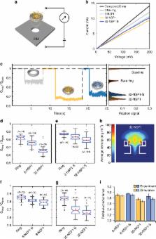
          The nuclear pore complex (NPC) is the gatekeeper for nuclear transport in eukaryotic cells. A key component of the NPC is the central shaft lined with intrinsically disordered proteins (IDPs) known as FG-Nups, which control the selective molecular traffic. Here, we present an approach to realize artificial NPC mimics that allows controlling the type and copy number of FG-Nups. We constructed 34 nm-wide 3D DNA origami rings and attached different numbers of NSP1, a model yeast FG-Nup, or NSP1-S, a hydrophilic mutant. Using (cryo) electron microscopy, we find that NSP1 forms denser cohesive networks inside the ring compared to NSP1-S. Consistent with this, the measured ionic conductance is lower for NSP1 than for NSP1-S. Molecular dynamics simulations reveal spatially varying protein densities and conductances in good agreement with the experiments. Our technique provides an experimental platform for deciphering the collective behavior of IDPs with full control of their type and position.

          Abstract

          FG-Nups are disordered proteins in the nuclear pore complex (NPC) where they selectively control nuclear transport. Here authors build NPC-mimics based on DNA origami rings which attach a certain numbers of Nups to analyse those nanopores by cryoEM and conductance measurements.

          Related collections

          Most cited references37

          • Record: found
          • Abstract: found
          • Article: not found

          Solid-state nanopores.

          The passage of individual molecules through nanosized pores in membranes is central to many processes in biology. Previously, experiments have been restricted to naturally occurring nanopores, but advances in technology now allow artificial solid-state nanopores to be fabricated in insulating membranes. By monitoring ion currents and forces as molecules pass through a solid-state nanopore, it is possible to investigate a wide range of phenomena involving DNA, RNA and proteins. The solid-state nanopore proves to be a surprisingly versatile new single-molecule tool for biophysics and biotechnology.
            Bookmark
            • Record: found
            • Abstract: found
            • Article: not found

            Folding DNA into twisted and curved nanoscale shapes.

            We demonstrate the ability to engineer complex shapes that twist and curve at the nanoscale from DNA. Through programmable self-assembly, strands of DNA are directed to form a custom-shaped bundle of tightly cross-linked double helices, arrayed in parallel to their helical axes. Targeted insertions and deletions of base pairs cause the DNA bundles to develop twist of either handedness or to curve. The degree of curvature could be quantitatively controlled, and a radius of curvature as tight as 6 nanometers was achieved. We also combined multiple curved elements to build several different types of intricate nanostructures, such as a wireframe beach ball or square-toothed gears.
              Bookmark
              • Record: found
              • Abstract: found
              • Article: found
              Is Open Access

              Rapid prototyping of 3D DNA-origami shapes with caDNAno

              DNA nanotechnology exploits the programmable specificity afforded by base-pairing to produce self-assembling macromolecular objects of custom shape. For building megadalton-scale DNA nanostructures, a long ‘scaffold’ strand can be employed to template the assembly of hundreds of oligonucleotide ‘staple’ strands into a planar antiparallel array of cross-linked helices. We recently adapted this ‘scaffolded DNA origami’ method to producing 3D shapes formed as pleated layers of double helices constrained to a honeycomb lattice. However, completing the required design steps can be cumbersome and time-consuming. Here we present caDNAno, an open-source software package with a graphical user interface that aids in the design of DNA sequences for folding 3D honeycomb-pleated shapes A series of rectangular-block motifs were designed, assembled, and analyzed to identify a well-behaved motif that could serve as a building block for future studies. The use of caDNAno significantly reduces the effort required to design 3D DNA-origami structures. The software is available at http://cadnano.org/, along with example designs and video tutorials demonstrating their construction. The source code is released under the MIT license.
                Bookmark

                Author and article information

                Contributors
                dietz@tum.de
                c.dekker@tudelft.nl
                Journal
                Nat Commun
                Nat Commun
                Nature Communications
                Nature Publishing Group UK (London )
                2041-1723
                2 March 2018
                2 March 2018
                2018
                : 9
                : 902
                Affiliations
                [1 ]ISNI 0000000123222966, GRID grid.6936.a, Physik Department and Institute for Advanced Study, , Technische Universität München, Am Coulombwall 4a, ; Garching bei München, D-85748 Germany
                [2 ]ISNI 0000 0001 2097 4740, GRID grid.5292.c, Department of Bionanoscience, Kavli Institute of Nanoscience, , Delft University of Technology, ; Van der Maasweg 9, 2629 HZ Delft, The Netherlands
                [3 ]ISNI 0000 0004 0407 1981, GRID grid.4830.f, Zernike Institute for Advanced Materials, , University of Groningen, Nijenborgh 4, ; 9747AG Groningen, The Netherlands
                Article
                3313
                10.1038/s41467-018-03313-w
                5834454
                29500415
                45c2ffbf-be73-4e04-b684-ad24204b2eff
                © The Author(s) 2018

                Open Access This article is licensed under a Creative Commons Attribution 4.0 International License, which permits use, sharing, adaptation, distribution and reproduction in any medium or format, as long as you give appropriate credit to the original author(s) and the source, provide a link to the Creative Commons license, and indicate if changes were made. The images or other third party material in this article are included in the article’s Creative Commons license, unless indicated otherwise in a credit line to the material. If material is not included in the article’s Creative Commons license and your intended use is not permitted by statutory regulation or exceeds the permitted use, you will need to obtain permission directly from the copyright holder. To view a copy of this license, visit http://creativecommons.org/licenses/by/4.0/.

                History
                : 6 November 2017
                : 2 February 2018
                Categories
                Article
                Custom metadata
                © The Author(s) 2018

                Uncategorized
                Uncategorized

                Comments

                Comment on this article