- Record: found
- Abstract: found
- Article: found
MTH1 Inhibitor TH1579 Induces Oxidative DNA Damage and Mitotic Arrest in Acute Myeloid Leukemia

Read this article at
Abstract
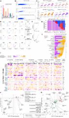
The MTH1 inhibitor TH1579 is a potential novel AML treatment, targeting both blasts and the pivotal leukemic stem cells while sparing normal bone marrow cells.
Abstract
Acute myeloid leukemia (AML) is an aggressive hematologic malignancy, exhibiting high levels of reactive oxygen species (ROS). ROS levels have been suggested to drive leukemogenesis and is thus a potential novel target for treating AML. MTH1 prevents incorporation of oxidized nucleotides into the DNA to maintain genome integrity and is upregulated in many cancers. Here we demonstrate that hematologic cancers are highly sensitive to MTH1 inhibitor TH1579 (karonudib). A functional precision medicine ex vivo screen in primary AML bone marrow samples demonstrated a broad response profile of TH1579, independent of the genomic alteration of AML, resembling the response profile of the standard-of-care treatments cytarabine and doxorubicin. Furthermore, TH1579 killed primary human AML blast cells (CD45 +) as well as chemotherapy resistance leukemic stem cells (CD45 +Lin −CD34 +CD38 −), which are often responsible for AML progression. TH1579 killed AML cells by causing mitotic arrest, elevating intracellular ROS levels, and enhancing oxidative DNA damage. TH1579 showed a significant therapeutic window, was well tolerated in animals, and could be combined with standard-of-care treatments to further improve efficacy. TH1579 significantly improved survival in two different AML disease models in vivo. In conclusion, the preclinical data presented here support that TH1579 is a promising novel anticancer agent for AML, providing a rationale to investigate the clinical usefulness of TH1579 in AML in an ongoing clinical phase I trial.
Related collections
Most cited references49
- Record: found
- Abstract: found
- Article: not found
Diagnosis and management of AML in adults: 2017 ELN recommendations from an international expert panel.
- Record: found
- Abstract: found
- Article: found
A Landscape of Pharmacogenomic Interactions in Cancer
- Record: found
- Abstract: found
- Article: not found