8
views
0
recommends
+1 Recommend
0 collections
    0
    shares
      • Record: found
      • Abstract: found
      • Article: found
      Is Open Access

      Characterization of Retinal Ganglion Cell and Optic Nerve Phenotypes Caused by Sustained Intracranial Pressure Elevation in Mice

      research-article

      Read this article at

      Bookmark
          There is no author summary for this article yet. Authors can add summaries to their articles on ScienceOpen to make them more accessible to a non-specialist audience.

          Abstract

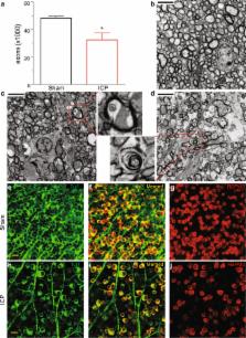
          Elevated intracranial pressure (ICP) can result in multiple neurologic sequelae including vision loss. Inducible models of ICP elevation are lacking in model organisms, which limits our understanding of the mechanism by which increased ICP impacts the visual system. We adapted a mouse model for the sustained elevation of ICP and tested the hypothesis that elevated ICP impacts the optic nerve and retinal ganglion cells (RGCs). ICP was elevated and maintained for 2 weeks, and resulted in multiple anatomic changes that are consistent with human disease including papilledema, loss of physiologic cupping, and engorgement of the optic nerve head. Elevated ICP caused a loss of RGC somas in the retina and RGC axons within the optic nerve, as well as a reduction in both RGC electrical function and contrast sensitivity. Elevated ICP also caused increased hypoxia-inducible factor (HIF)-1 alpha expression in the ganglion cell layer. These experiments confirm that sustained ICP elevation can be achieved in mice and causes phenotypes that preferentially impact RGCs and are similar to those seen in human disease. With this model, it is possible to model human diseases of elevated ICP such as Idiopathic Intracranial Hypertension and Spaceflight Associated Neuro-ocular Syndrome.

          Related collections

          Most cited references53

          • Record: found
          • Abstract: found
          • Article: not found

          Cerebrospinal fluid pressure in glaucoma: a prospective study.

          To assess whether a low cerebrospinal fluid pressure (CSF-P) is associated with open-angle glaucoma in eyes with normal intraocular pressure (IOP). Prospective, interventional study. The study included 43 patients with open-angle glaucoma (14 with a normal IOP, and 29 with an elevated IOP) and 71 subjects without glaucoma. All patients underwent standardized ophthalmologic and neurologic examinations and measurement of lumbar CSF-P. Cerebrospinal fluid pressure and IOP. Lumbar CSF-P was significantly (P<0.001) lower in the normal IOP glaucoma group (9.5+/-2.2 mmHg) than in the high IOP glaucoma group (11.7+/-2.7 mmHg) or the control group (12.9+/-1.9 mmHg). The trans-lamina cribrosa pressure difference (IOP minus CSF-P) was significantly (P<0.001) higher in the normal IOP glaucoma group (6.6+/-3.6 mmHg) and the high-IOP glaucoma group (12.5+/-4.1 mmHg) than in the control group (1.4+/-1.7 mmHg). The extent of glaucomatous visual field loss was negatively correlated with the height of the CSF-P and positively correlated with the trans-lamina cribrosa pressure difference. In the control group, CSF-P was significantly correlated with both systolic blood pressure (P = 0.04) and IOP (P<0.001). The trans-lamina cribrosa pressure difference was not significantly associated with blood pressure (P = 0.97). In open-angle glaucoma with normal IOP, CSF-P is abnormally low, leading to an abnormally high trans-lamina cribrosa pressure difference. Pathogenetically, a low CSF-P in normal-IOP glaucoma may be similar to a high IOP in high-IOP glaucoma. Consequently, the glaucomatous visual field defect is positively correlated with the trans-lamina cribrosa pressure difference and inversely correlated with the CSF-P. In nonglaucomatous subjects, CSF-P, blood pressure, and IOP are significantly associated with each other. Copyright (c) 2010 American Academy of Ophthalmology. Published by Elsevier Inc. All rights reserved.
            Bookmark
            • Record: found
            • Abstract: found
            • Article: not found

            'Hit & Run' model of closed-skull traumatic brain injury (TBI) reveals complex patterns of post-traumatic AQP4 dysregulation.

            Cerebral edema is a major contributor to morbidity associated with traumatic brain injury (TBI). The methods involved in most rodent models of TBI, including head fixation, opening of the skull, and prolonged anesthesia, likely alter TBI development and reduce secondary injury. We report the development of a closed-skull model of murine TBI, which minimizes time of anesthesia, allows the monitoring of intracranial pressure (ICP), and can be modulated to produce mild and moderate grade TBI. In this model, we characterized changes in aquaporin-4 (AQP4) expression and localization after mild and moderate TBI. We found that global AQP4 expression after TBI was generally increased; however, analysis of AQP4 localization revealed that the most prominent effect of TBI on AQP4 was the loss of polarized localization at endfoot processes of reactive astrocytes. This AQP4 dysregulation peaked at 7 days after injury and was largely indistinguishable between mild and moderate grade TBI for the first 2 weeks after injury. Within the same model, blood-brain barrieranalysis of variance permeability, cerebral edema, and ICP largely normalized within 7 days after moderate TBI. These findings suggest that changes in AQP4 expression and localization may not contribute to cerebral edema formation, but rather may represent a compensatory mechanism to facilitate its resolution.
              Bookmark
              • Record: found
              • Abstract: found
              • Article: not found

              Cerebrospinal fluid pressure is decreased in primary open-angle glaucoma.

              To compare cerebrospinal fluid (CSF) pressure in patients with primary open-angle glaucoma (POAG) with that in nonglaucomatous patients. Case-control study. Thirty-one thousand, seven hundred and eighty-six subjects underwent lumbar puncture (LP) between 1996 and 2007 at the Mayo Clinic, Rochester, Minnesota. Of these, 28 patients who had POAG and 49 patients who did not have POAG were analyzed. Retrospective review of medical records. Comparison of the 2 groups and factors associated with CSF pressure were analyzed by univariate and multivariate analyses. Demographics (age and gender), medical history, medication use, indication for LP, intraocular pressure (IOP), optic disc cup-to-disc ratio, visual field assessment, and CSF pressure. The mean CSF pressure +/- standard deviation was 13.0+/-4.2 mmHg in nonglaucoma patients and 9.2+/-2.9 mmHg in POAG patients (P<0.00005). The CSF pressure was lower in POAG patients regardless of indication for LP or age. Linear regression analysis showed that cup-to-disc ratio correlated independently with IOP (P<0.0001), CSF pressure (P<0.0001), and the translaminar pressure difference (P<0.0001). Multivariate analysis demonstrated that larger cup-to-disc ratio (P<0.0001) was associated with lower CSF pressure. Cerebrospinal fluid pressure is significantly lower in POAG patients compared with that in nonglaucomatous controls. These data support the notion that CSF pressure may play an important contributory role in the pathogenesis of POAG.
                Bookmark

                Author and article information

                Contributors
                benjamin.frankfort@bcm.edu
                Journal
                Sci Rep
                Sci Rep
                Scientific Reports
                Nature Publishing Group UK (London )
                2045-2322
                12 February 2018
                12 February 2018
                2018
                : 8
                : 2856
                Affiliations
                [1 ]ISNI 0000 0001 2160 926X, GRID grid.39382.33, Department of Ophthalmology, , Baylor College of Medicine, ; Houston, TX USA
                [2 ]ISNI 0000 0001 2160 926X, GRID grid.39382.33, Department of Neuroscience, , Baylor College of Medicine, ; Houston, TX USA
                [3 ]ISNI 0000 0004 1764 6123, GRID grid.16890.36, School of Optometry, , The Hong Kong Polytechnic University, ; Hong Kong, Hong Kong
                Author information
                http://orcid.org/0000-0001-7561-9121
                Article
                21254
                10.1038/s41598-018-21254-8
                5809383
                29434244
                490d6668-a2ab-498e-95ef-8669d719d491
                © The Author(s) 2018

                Open Access This article is licensed under a Creative Commons Attribution 4.0 International License, which permits use, sharing, adaptation, distribution and reproduction in any medium or format, as long as you give appropriate credit to the original author(s) and the source, provide a link to the Creative Commons license, and indicate if changes were made. The images or other third party material in this article are included in the article’s Creative Commons license, unless indicated otherwise in a credit line to the material. If material is not included in the article’s Creative Commons license and your intended use is not permitted by statutory regulation or exceeds the permitted use, you will need to obtain permission directly from the copyright holder. To view a copy of this license, visit http://creativecommons.org/licenses/by/4.0/.

                History
                : 10 November 2017
                : 31 January 2018
                Categories
                Article
                Custom metadata
                © The Author(s) 2018

                Uncategorized
                Uncategorized

                Comments

                Comment on this article