29
views
0
recommends
+1 Recommend
0 collections
    0
    shares
      • Record: found
      • Abstract: found
      • Article: found
      Is Open Access

      CRISPR screening using an expanded toolkit of autophagy reporters identifies TMEM41B as a novel autophagy factor

      research-article

      Read this article at

      Bookmark
          There is no author summary for this article yet. Authors can add summaries to their articles on ScienceOpen to make them more accessible to a non-specialist audience.

          Abstract

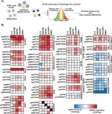
          The power of forward genetics in yeast is the foundation on which the field of autophagy research firmly stands. Complementary work on autophagy in higher eukaryotes has revealed both the deep conservation of this process, as well as novel mechanisms by which autophagy is regulated in the context of development, immunity, and neuronal homeostasis. The recent emergence of new clustered regularly interspaced palindromic repeats/CRISPR-associated protein 9 (CRISPR/Cas9)-based technologies has begun facilitating efforts to define novel autophagy factors and pathways by forward genetic screening in mammalian cells. Here, we set out to develop an expanded toolkit of autophagy reporters amenable to CRISPR/Cas9 screening. Genome-wide screening of our reporters in mammalian cells recovered virtually all known autophagy-related (ATG) factors as well as previously uncharacterized factors, including vacuolar protein sorting 37 homolog A (VPS37A), transmembrane protein 251 (TMEM251), amyotrophic lateral sclerosis 2 (ALS2), and TMEM41B. To validate this data set, we used quantitative microscopy and biochemical analyses to show that 1 novel hit, TMEM41B, is required for phagophore maturation. TMEM41B is an integral endoplasmic reticulum (ER) membrane protein distantly related to the established autophagy factor vacuole membrane protein 1 (VMP1), and our data show that these two factors play related, albeit not fully overlapping, roles in autophagosome biogenesis. In sum, our work uncovers new ATG factors, reveals a malleable network of autophagy receptor genetic interactions, and provides a valuable resource ( http://crispr.deniclab.com) for further mining of novel autophagy mechanisms.

          Author summary

          Eukaryotic cells use autophagy to eliminate unwanted structures—such as protein aggregates, intracellular pathogens, and damaged organelles—that are too large to be handled by the proteasome. This unusual vesicle transport pathway begins with packaging of cytoplasmic targets into a double-membrane vesicle (autophagosome) and ends with their degradation in the lysosome. A deeper understanding of autophagy as a regulated mechanism for protein turnover has the potential to provide new therapies for diverse human pathologies, including neurodegenerative disease, cardiometabolic disease, and cancer. Here, we set out to build new reporters for studying mammalian autophagy by genetic screening. This approach allowed us to carry out pooled genome-wide clustered regularly interspaced palindromic repeats (CRISPR) knockout screens and recover virtually all known mammalian autophagy-related (ATG) factors. In addition, we uncovered several uncharacterized proteins, including the endoplasmic reticulum (ER) transmembrane protein 41B (TMEM41B), which we went on to show is required for normal growth of autophagosome precursor membranes around their targets. More broadly, our data set provides a comprehensive resource of genes that affect autophagosome trafficking to the lysosome for further hypothesis testing.

          Related collections

          Most cited references48

          • Record: found
          • Abstract: found
          • Article: not found

          Dynamics and diversity in autophagy mechanisms: lessons from yeast.

          Autophagy is a fundamental function of eukaryotic cells and is well conserved from yeast to humans. The most remarkable feature of autophagy is the synthesis of double membrane-bound compartments that sequester materials to be degraded in lytic compartments, a process that seems to be mechanistically distinct from conventional membrane traffic. The discovery of autophagy in yeast and the genetic tractability of this organism have allowed us to identify genes that are responsible for this process, which has led to the explosive growth of this research field seen today. Analyses of autophagy-related (Atg) proteins have unveiled dynamic and diverse aspects of mechanisms that underlie membrane formation during autophagy.
            Bookmark
            • Record: found
            • Abstract: found
            • Article: not found

            Discovery of Atg5/Atg7-independent alternative macroautophagy.

            Macroautophagy is a process that leads to the bulk degradation of subcellular constituents by producing autophagosomes/autolysosomes. It is believed that Atg5 (ref. 4) and Atg7 (ref. 5) are essential genes for mammalian macroautophagy. Here we show, however, that mouse cells lacking Atg5 or Atg7 can still form autophagosomes/autolysosomes and perform autophagy-mediated protein degradation when subjected to certain stressors. Although lipidation of the microtubule-associated protein light chain 3 (LC3, also known as Map1lc3a) to form LC3-II is generally considered to be a good indicator of macroautophagy, it did not occur during the Atg5/Atg7-independent alternative process of macroautophagy. We also found that this alternative process of macroautophagy was regulated by several autophagic proteins, including Unc-51-like kinase 1 (Ulk1) and beclin 1. Unlike conventional macroautophagy, autophagosomes seemed to be generated in a Rab9-dependent manner by the fusion of isolation membranes with vesicles derived from the trans-Golgi and late endosomes. In vivo, Atg5-independent alternative macroautophagy was detected in several embryonic tissues. It also had a function in clearing mitochondria during erythroid maturation. These results indicate that mammalian macroautophagy can occur through at least two different pathways: an Atg5/Atg7-dependent conventional pathway and an Atg5/Atg7-independent alternative pathway.
              Bookmark
              • Record: found
              • Abstract: found
              • Article: found
              Is Open Access

              Atg8 family LC3/GABARAP proteins are crucial for autophagosome–lysosome fusion but not autophagosome formation during PINK1/Parkin mitophagy and starvation

              Current autophagy models suggest that Atg8 family LC3/GABARAP proteins are essential mediators of autophagosome biogenesis. Nguyen et al. exploit CRISPR/Cas9-generated knockouts of the LC3 or GABARAP subfamilies, or both subfamilies, to show that Atg8s are dispensable for autophagosome biogenesis but essential for autophagosome–lysosome fusion.
                Bookmark

                Author and article information

                Contributors
                Role: ConceptualizationRole: Formal analysisRole: Funding acquisitionRole: InvestigationRole: MethodologyRole: ValidationRole: VisualizationRole: Writing – original draftRole: Writing – review & editing
                Role: InvestigationRole: Methodology
                Role: Data curationRole: Formal analysisRole: Visualization
                Role: InvestigationRole: Resources
                Role: Formal analysisRole: InvestigationRole: Methodology
                Role: ConceptualizationRole: Funding acquisitionRole: SupervisionRole: Writing – review & editing
                Role: Academic Editor
                Journal
                PLoS Biol
                PLoS Biol
                plos
                plosbiol
                PLoS Biology
                Public Library of Science (San Francisco, CA USA )
                1544-9173
                1545-7885
                1 April 2019
                April 2019
                1 April 2019
                : 17
                : 4
                : e2007044
                Affiliations
                [1 ] Department of Molecular and Cellular Biology, Harvard University, Northwest Labs, Cambridge, Massachusetts, United States of America
                [2 ] Centre for Cancer Cell Reprogramming, Faculty of Medicine, University of Oslo, Oslo, Norway
                [3 ] Department of Molecular Cell Biology, Institute for Cancer Research, Oslo University Hospital, Oslo, Norway
                University of Oslo, Norway
                Author notes

                The authors have declared that no competing interests exist.

                [¤]

                Current address: Department of Biochemistry and Cell Biology, Geisel School of Medicine at Dartmouth, Hanover, New Hampshire, United States of America

                Author information
                http://orcid.org/0000-0002-1982-7281
                Article
                pbio.2007044
                10.1371/journal.pbio.2007044
                6459555
                30933966
                4b1790e2-90ac-427a-9528-ca4a2626c4ee
                © 2019 Shoemaker et al

                This is an open access article distributed under the terms of the Creative Commons Attribution License, which permits unrestricted use, distribution, and reproduction in any medium, provided the original author and source are credited.

                History
                : 20 June 2018
                : 13 March 2019
                Page count
                Figures: 8, Tables: 0, Pages: 31
                Funding
                National Institutes of Health (grant number 1R35GM127136) to VD. The funder had no role in study design, data collection and analysis, decision to publish, or preparation of the manuscript. National Institutes of Health (grant number 5K99GM117218) to CJS. The funder had no role in study design, data collection and analysis, decision to publish, or preparation of the manuscript. National Institutes of Health (grant number P20-GM113132). The funder had no role in study design, data collection and analysis, decision to publish, or preparation of the manuscript.
                Categories
                Methods and Resources
                Biology and Life Sciences
                Cell Biology
                Cell Processes
                Cell Death
                Autophagic Cell Death
                Biology and Life Sciences
                Biochemistry
                Proteins
                Luminescent Proteins
                Green Fluorescent Protein
                Biology and Life Sciences
                Cell Biology
                Cellular Structures and Organelles
                Cell Membranes
                Membrane Proteins
                Biology and Life Sciences
                Cell Biology
                Cellular Structures and Organelles
                Vesicles
                Research and Analysis Methods
                Spectrum Analysis Techniques
                Spectrophotometry
                Cytophotometry
                Flow Cytometry
                Biology and Life Sciences
                Bioengineering
                Synthetic Bioengineering
                Genome Engineering
                Synthetic Genome Editing
                Crispr
                Engineering and Technology
                Bioengineering
                Synthetic Bioengineering
                Genome Engineering
                Synthetic Genome Editing
                Crispr
                Biology and Life Sciences
                Synthetic Biology
                Synthetic Bioengineering
                Genome Engineering
                Synthetic Genome Editing
                Crispr
                Engineering and Technology
                Synthetic Biology
                Synthetic Bioengineering
                Genome Engineering
                Synthetic Genome Editing
                Crispr
                Biology and Life Sciences
                Synthetic Biology
                Synthetic Genomics
                Synthetic Genome Editing
                Crispr
                Engineering and Technology
                Synthetic Biology
                Synthetic Genomics
                Synthetic Genome Editing
                Crispr
                Biology and Life Sciences
                Biochemistry
                Biosynthesis
                Biology and Life Sciences
                Biochemistry
                Lipids
                Custom metadata
                vor-update-to-uncorrected-proof
                2019-04-11
                All relevant data are within the paper and its Supporting Information files.

                Life sciences
                Life sciences

                Comments

                Comment on this article