56
views
0
recommends
+1 Recommend
0 collections
    0
    shares
      • Record: found
      • Abstract: found
      • Article: found
      Is Open Access

      Atg8 family LC3/GABARAP proteins are crucial for autophagosome–lysosome fusion but not autophagosome formation during PINK1/Parkin mitophagy and starvation

      research-article

      Read this article at

      Bookmark
          There is no author summary for this article yet. Authors can add summaries to their articles on ScienceOpen to make them more accessible to a non-specialist audience.

          Abstract

          Current autophagy models suggest that Atg8 family LC3/GABARAP proteins are essential mediators of autophagosome biogenesis. Nguyen et al. exploit CRISPR/Cas9-generated knockouts of the LC3 or GABARAP subfamilies, or both subfamilies, to show that Atg8s are dispensable for autophagosome biogenesis but essential for autophagosome–lysosome fusion.

          Abstract

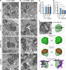
          Members of the Atg8 family of proteins are conjugated to autophagosomal membranes, where they have been proposed to drive autophagosome formation and selective sequestration of cargo. In mammals, the Atg8 family consists of six members divided into the LC3 and GABARAP subfamilies. To define Atg8 function, we used genome editing to generate knockouts of the LC3 and GABARAP subfamilies as well as all six Atg8 family members in HeLa cells. We show that Atg8s are dispensable for autophagosome formation and selective engulfment of mitochondria, but essential for autophagosome–lysosome fusion. We find that the GABARAP subfamily promotes PLEKHM1 recruitment and governs autophagosome–lysosome fusion, whereas the LC3 subfamily plays a less prominent role in these processes. Although neither GABARAPs nor LC3s are required for autophagosome biogenesis, loss of all Atg8s yields smaller autophagosomes and a slowed initial rate of autophagosome formation. Our results clarify the essential function of the Atg8 family and identify GABARAP subfamily members as primary contributors to PINK1/Parkin mitophagy and starvation autophagy.

          Related collections

          Most cited references29

          • Record: found
          • Abstract: found
          • Article: found
          Is Open Access

          Autophagosome formation from membrane compartments enriched in phosphatidylinositol 3-phosphate and dynamically connected to the endoplasmic reticulum

          Autophagy is the engulfment of cytosol and organelles by double-membrane vesicles termed autophagosomes. Autophagosome formation is known to require phosphatidylinositol 3-phosphate (PI(3)P) and occurs near the endoplasmic reticulum (ER), but the exact mechanisms are unknown. We show that double FYVE domain–containing protein 1, a PI(3)P-binding protein with unusual localization on ER and Golgi membranes, translocates in response to amino acid starvation to a punctate compartment partially colocalized with autophagosomal proteins. Translocation is dependent on Vps34 and beclin function. Other PI(3)P-binding probes targeted to the ER show the same starvation-induced translocation that is dependent on PI(3)P formation and recognition. Live imaging experiments show that this punctate compartment forms near Vps34-containing vesicles, is in dynamic equilibrium with the ER, and provides a membrane platform for accumulation of autophagosomal proteins, expansion of autophagosomal membranes, and emergence of fully formed autophagosomes. This PI(3)P-enriched compartment may be involved in autophagosome biogenesis. Its dynamic relationship with the ER is consistent with the idea that the ER may provide important components for autophagosome formation.
            Bookmark
            • Record: found
            • Abstract: found
            • Article: not found

            A ubiquitin-like system mediates protein lipidation.

            Autophagy is a dynamic membrane phenomenon for bulk protein degradation in the lysosome/vacuole. Apg8/Aut7 is an essential factor for autophagy in yeast. We previously found that the carboxy-terminal arginine of nascent Apg8 is removed by Apg4/Aut2 protease, leaving a glycine residue at the C terminus. Apg8 is then converted to a form (Apg8-X) that is tightly bound to the membrane. Here we report a new mode of protein lipidation. Apg8 is covalently conjugated to phosphatidylethanolamine through an amide bond between the C-terminal glycine and the amino group of phosphatidylethanolamine. This lipidation is mediated by a ubiquitination-like system. Apg8 is a ubiquitin-like protein that is activated by an E1 protein, Apg7 (refs 7, 8), and is transferred subsequently to the E2 enzymes Apg3/Aut1 (ref. 9). Apg7 activates two different ubiquitin-like proteins, Apg12 (ref. 10) and Apg8, and assigns them to specific E2 enzymes, Apg10 (ref. 11) and Apg3, respectively. These reactions are necessary for the formation of Apg8-phosphatidylethanolamine. This lipidation has an essential role in membrane dynamics during autophagy.
              Bookmark
              • Record: found
              • Abstract: found
              • Article: not found

              Nix is a selective autophagy receptor for mitochondrial clearance.

              Autophagy is the cellular homeostatic pathway that delivers large cytosolic materials for degradation in the lysosome. Recent evidence indicates that autophagy mediates selective removal of protein aggregates, organelles and microbes in cells. Yet, the specificity in targeting a particular substrate to the autophagy pathway remains poorly understood. Here, we show that the mitochondrial protein Nix is a selective autophagy receptor by binding to LC3/GABARAP proteins, ubiquitin-like modifiers that are required for the growth of autophagosomal membranes. In cultured cells, Nix recruits GABARAP-L1 to damaged mitochondria through its amino-terminal LC3-interacting region. Furthermore, ablation of the Nix:LC3/GABARAP interaction retards mitochondrial clearance in maturing murine reticulocytes. Thus, Nix functions as an autophagy receptor, which mediates mitochondrial clearance after mitochondrial damage and during erythrocyte differentiation.
                Bookmark

                Author and article information

                Journal
                J Cell Biol
                J. Cell Biol
                jcb
                jcb
                The Journal of Cell Biology
                The Rockefeller University Press
                0021-9525
                1540-8140
                19 December 2016
                : 215
                : 6
                : 857-874
                Affiliations
                [1 ]Department of Biochemistry and Molecular Biology, Biomedicine Discovery Institute, Monash University, Melbourne, Victoria 3800, Australia
                [2 ]Monash Ramaciotti Centre for Cryo Electron Microscopy, Monash University, Melbourne, Victoria 3800, Australia
                Author notes
                Correspondence to Michael Lazarou: michael.lazarou@ 123456monash.edu
                [*]

                T.N. Nguyen and B.S. Padman contributed equally to this paper.

                Author information
                http://orcid.org/0000-0001-9698-0020
                http://orcid.org/0000-0002-5710-6100
                http://orcid.org/0000-0002-4926-802X
                http://orcid.org/0000-0002-8122-8271
                http://orcid.org/0000-0003-3596-2288
                http://orcid.org/0000-0003-2150-5545
                Article
                201607039
                10.1083/jcb.201607039
                5166504
                27864321
                3f79996c-dee2-43b6-9e3e-213138502842
                © 2016 Nguyen et al.

                This article is distributed under the terms of an Attribution–Noncommercial–Share Alike–No Mirror Sites license for the first six months after the publication date (see http://www.rupress.org/terms). After six months it is available under a Creative Commons License (Attribution–Noncommercial–Share Alike 3.0 Unported license, as described at http://creativecommons.org/licenses/by-nc-sa/3.0/).

                History
                : 12 July 2016
                : 06 October 2016
                : 09 November 2016
                Funding
                Funded by: National Health and Medical Research Council https://doi.org/10.13039/501100000925
                Award ID: GNT1063781
                Award ID: GNT1106471
                Categories
                Research Articles
                Article
                41
                19
                22

                Cell biology
                Cell biology

                Comments

                Comment on this article