1
views
0
recommends
+1 Recommend
0 collections
    0
    shares
      • Record: found
      • Abstract: found
      • Article: found
      Is Open Access

      In vivo quantitative photoacoustic evaluation of the liver and kidney pathology in tyrosinemia

      research-article

      Read this article at

      Bookmark
          There is no author summary for this article yet. Authors can add summaries to their articles on ScienceOpen to make them more accessible to a non-specialist audience.

          Abstract

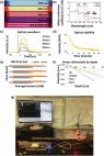
          Hereditary tyrosinemia type Ⅰ (HT1) is a severe autosomal recessive inherited metabolic disease, which can result in severe damage of liver and kidney. Photoacoustic imaging (PAI) uses pulsed laser light to induce ultrasonic signals to facilitate the visualization of lesions that are strongly related to disease progression. In this study, the structural and functional changes of liver and kidney in HT1 was investigated by cross-scale PAI. The results showed that the hepatic lobule and renal tubule were severely damaged during HT1 progression. The hemoglobin content, vessel density, and liver function reserve were decreased. The metabolic half-life of indocyanine green declined from 59.8 s in health to 262.6 s in the advanced stage. Blood oxygen saturation was much lower than that in health. This study highlights the potential of PAI for in vivo evaluation of the liver and kidney lesions in HT1.

          Related collections

          Most cited references50

          • Record: found
          • Abstract: found
          • Article: not found

          Contrast agents for molecular photoacoustic imaging.

          Photoacoustic imaging (PAI) is an emerging tool that bridges the traditional depth limits of ballistic optical imaging and the resolution limits of diffuse optical imaging. Using the acoustic waves generated in response to the absorption of pulsed laser light, it provides noninvasive images of absorbed optical energy density at depths of several centimeters with a resolution of ∼100 μm. This versatile and scalable imaging modality has now shown potential for molecular imaging, which enables visualization of biological processes with systemically introduced contrast agents. Understanding the relative merits of the vast range of contrast agents available, from small-molecule dyes to gold and carbon nanostructures to liposome encapsulations, is a considerable challenge. Here we critically review the physical, chemical and biochemical characteristics of the existing photoacoustic contrast agents, highlighting key applications and present challenges for molecular PAI.
            Bookmark
            • Record: found
            • Abstract: found
            • Article: not found

            A practical guide to photoacoustic tomography in the life sciences.

            The life sciences can benefit greatly from imaging technologies that connect microscopic discoveries with macroscopic observations. One technology uniquely positioned to provide such benefits is photoacoustic tomography (PAT), a sensitive modality for imaging optical absorption contrast over a range of spatial scales at high speed. In PAT, endogenous contrast reveals a tissue's anatomical, functional, metabolic, and histologic properties, and exogenous contrast provides molecular and cellular specificity. The spatial scale of PAT covers organelles, cells, tissues, organs, and small animals. Consequently, PAT is complementary to other imaging modalities in contrast mechanism, penetration, spatial resolution, and temporal resolution. We review the fundamentals of PAT and provide practical guidelines for matching PAT systems with research needs. We also summarize the most promising biomedical applications of PAT, discuss related challenges, and envision PAT's potential to lead to further breakthroughs.
              Bookmark
              • Record: found
              • Abstract: found
              • Article: found
              Is Open Access

              A review of clinical photoacoustic imaging: Current and future trends

              Photoacoustic imaging (or optoacoustic imaging) is an upcoming biomedical imaging modality availing the benefits of optical resolution and acoustic depth of penetration. With its capacity to offer structural, functional, molecular and kinetic information making use of either endogenous contrast agents like hemoglobin, lipid, melanin and water or a variety of exogenous contrast agents or both, PAI has demonstrated promising potential in a wide range of preclinical and clinical applications. This review provides an overview of the rapidly expanding clinical applications of photoacoustic imaging including breast imaging, dermatologic imaging, vascular imaging, carotid artery imaging, musculoskeletal imaging, gastrointestinal imaging and adipose tissue imaging and the future directives utilizing different configurations of photoacoustic imaging. Particular emphasis is placed on investigations performed on human or human specimens.
                Bookmark

                Author and article information

                Contributors
                Journal
                Photoacoustics
                Photoacoustics
                Photoacoustics
                Elsevier
                2213-5979
                28 September 2022
                December 2022
                28 September 2022
                : 28
                : 100410
                Affiliations
                [a ]Research Center of Medical Sciences, Guangdong Provincial People's Hospital, Guangdong Academy of Medical Sciences, 510000 Guangzhou, China
                [b ]Guangdong Cardiovsacular Institute, 510000 Guangzhou, China
                [c ]School of Public Health, Xiamen University, 361005 Xiamen, China
                [d ]State Key Laboratory of Precision Electronic Manufacturing Technology and Equipment, Guangdong University of Technology, 510000 Guangzhou, China
                [e ]Department of Hepatobiliary Surgery, Zhujiang Hospital, Southern Medical University, 510000 Guangzhou, China
                [f ]Guangdong Provincial Key Laboratory of Artificial Intelligence in Medical Image Analysis and Application, Guangdong Provincial People's Hospital, Guangdong Academy of Medical Sciences, 510000 Guangzhou, China
                Author notes
                [* ]Corresponding author. zenglvming@ 123456163.com
                [** ]Corresponding author at: Research Center of Medical Sciences, Guangdong Provincial People's Hospital, Guangdong Academy of Medical Sciences, 510000 Guangzhou, China. limingnie@ 123456gmail.com
                [1]

                These authors contributed equally.

                Article
                S2213-5979(22)00075-1 100410
                10.1016/j.pacs.2022.100410
                9531282
                36204180
                4b50ac26-d85e-4182-9fb8-a30bb1d49ec5
                © 2022 The Authors

                This is an open access article under the CC BY-NC-ND license (http://creativecommons.org/licenses/by-nc-nd/4.0/).

                History
                : 24 June 2022
                : 9 September 2022
                : 27 September 2022
                Categories
                Research Article

                photoacoustic imaging,tyrosinemia,hepatic lobule,renal tubule

                Comments

                Comment on this article