2
views
0
recommends
+1 Recommend
1 collections
    0
    shares
      • Record: found
      • Abstract: found
      • Article: not found

      Mechanistic insights of host cell fusion of SARS-CoV-1 and SARS-CoV-2 from atomic resolution structure and membrane dynamics

      review-article

      Read this article at

      ScienceOpenPublisherPMC
      Bookmark
          There is no author summary for this article yet. Authors can add summaries to their articles on ScienceOpen to make them more accessible to a non-specialist audience.

          Abstract

          The emerging and re-emerging viral diseases are continuous threats to the wellbeing of human life. Previous outbreaks of Severe Acute Respiratory Syndrome (SARS) and Middle East Respiratory Syndrome (MERS had evidenced potential threats of coronaviruses in human health. The recent pandemic due to SARS-CoV-2 is overwhelming and has been going beyond control. Vaccines and antiviral drugs are ungently required to mitigate the pandemic. Therefore, it is important to comprehend the mechanistic details of viral infection process. The fusion between host cell and virus being the first step of infection, understanding the fusion mechanism could provide crucial information to intervene the infection process. Interestingly, all enveloped viruses contain fusion protein on their envelope that acts as fusion machine. For coronaviruses, the spike or S glycoprotein mediates successful infection through receptor binding and cell fusion. The cell fusion process requires merging of virus and host cell membranes, and that is essentially performed by the S2 domain of the S glycoprotein. In this review, we have discussed cell fusion mechanism of SARS-CoV-1 from available atomic resolution structures and membrane binding of fusion peptides. We have further discussed about the cell fusion of SARS-CoV-2 in the context of present pandemic situation.
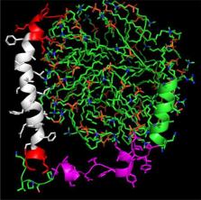
          Graphical abstract

          Highlights

          • Virus-host cell fusion is essential for infection.

          • Viral fusion proteins are involved in the process.

          • SARS-CoV-1 and SARS-CoV-2 fusion systems are reviewed.

          Related collections

          Most cited references93

          • Record: found
          • Abstract: found
          • Article: not found

          SARS-CoV-2 Cell Entry Depends on ACE2 and TMPRSS2 and Is Blocked by a Clinically Proven Protease Inhibitor

          Summary The recent emergence of the novel, pathogenic SARS-coronavirus 2 (SARS-CoV-2) in China and its rapid national and international spread pose a global health emergency. Cell entry of coronaviruses depends on binding of the viral spike (S) proteins to cellular receptors and on S protein priming by host cell proteases. Unravelling which cellular factors are used by SARS-CoV-2 for entry might provide insights into viral transmission and reveal therapeutic targets. Here, we demonstrate that SARS-CoV-2 uses the SARS-CoV receptor ACE2 for entry and the serine protease TMPRSS2 for S protein priming. A TMPRSS2 inhibitor approved for clinical use blocked entry and might constitute a treatment option. Finally, we show that the sera from convalescent SARS patients cross-neutralized SARS-2-S-driven entry. Our results reveal important commonalities between SARS-CoV-2 and SARS-CoV infection and identify a potential target for antiviral intervention.
            Bookmark
            • Record: found
            • Abstract: found
            • Article: found
            Is Open Access

            Cryo-EM structure of the 2019-nCoV spike in the prefusion conformation

            Structure of the nCoV trimeric spike The World Health Organization has declared the outbreak of a novel coronavirus (2019-nCoV) to be a public health emergency of international concern. The virus binds to host cells through its trimeric spike glycoprotein, making this protein a key target for potential therapies and diagnostics. Wrapp et al. determined a 3.5-angstrom-resolution structure of the 2019-nCoV trimeric spike protein by cryo–electron microscopy. Using biophysical assays, the authors show that this protein binds at least 10 times more tightly than the corresponding spike protein of severe acute respiratory syndrome (SARS)–CoV to their common host cell receptor. They also tested three antibodies known to bind to the SARS-CoV spike protein but did not detect binding to the 2019-nCoV spike protein. These studies provide valuable information to guide the development of medical counter-measures for 2019-nCoV. Science, this issue p. 1260
              Bookmark
              • Record: found
              • Abstract: found
              • Article: not found

              Structure, Function, and Antigenicity of the SARS-CoV-2 Spike Glycoprotein

              Summary The emergence of SARS-CoV-2 has resulted in >90,000 infections and >3,000 deaths. Coronavirus spike (S) glycoproteins promote entry into cells and are the main target of antibodies. We show that SARS-CoV-2 S uses ACE2 to enter cells and that the receptor-binding domains of SARS-CoV-2 S and SARS-CoV S bind with similar affinities to human ACE2, correlating with the efficient spread of SARS-CoV-2 among humans. We found that the SARS-CoV-2 S glycoprotein harbors a furin cleavage site at the boundary between the S1/S2 subunits, which is processed during biogenesis and sets this virus apart from SARS-CoV and SARS-related CoVs. We determined cryo-EM structures of the SARS-CoV-2 S ectodomain trimer, providing a blueprint for the design of vaccines and inhibitors of viral entry. Finally, we demonstrate that SARS-CoV S murine polyclonal antibodies potently inhibited SARS-CoV-2 S mediated entry into cells, indicating that cross-neutralizing antibodies targeting conserved S epitopes can be elicited upon vaccination.
                Bookmark

                Author and article information

                Contributors
                Journal
                Biophys Chem
                Biophys. Chem
                Biophysical Chemistry
                Published by Elsevier B.V.
                0301-4622
                1873-4200
                22 July 2020
                22 July 2020
                : 106438
                Affiliations
                [a ]School of Chemistry, Sambalpur University, Jyoti Vihar, Burla, Odisha 768 019, India
                [b ]Centre of Excellence in Natural Products and Therapeutics, Sambalpur University, Jyoti Vihar, Burla, Odisha 768 019, India
                [c ]School of Biological Sciences, Nanyang Technological University, 60 Nanyang Drive, Singapore 637551, Singapore
                Author notes
                [* ]Corresponding author. surajit@ 123456ntu.edu.sg
                Article
                S0301-4622(20)30146-0 106438
                10.1016/j.bpc.2020.106438
                7375304
                50872feb-82fd-47db-a630-25ab0a7e7ec3
                © 2020 Published by Elsevier B.V.

                Since January 2020 Elsevier has created a COVID-19 resource centre with free information in English and Mandarin on the novel coronavirus COVID-19. The COVID-19 resource centre is hosted on Elsevier Connect, the company's public news and information website. Elsevier hereby grants permission to make all its COVID-19-related research that is available on the COVID-19 resource centre - including this research content - immediately available in PubMed Central and other publicly funded repositories, such as the WHO COVID database with rights for unrestricted research re-use and analyses in any form or by any means with acknowledgement of the original source. These permissions are granted for free by Elsevier for as long as the COVID-19 resource centre remains active.

                History
                : 12 June 2020
                : 16 July 2020
                : 17 July 2020
                Categories
                Article

                Biochemistry
                sars-cov-1,sars-cov-2,covid-19,fusion peptide,fusion protein,cell fusion
                Biochemistry
                sars-cov-1, sars-cov-2, covid-19, fusion peptide, fusion protein, cell fusion

                Comments

                Comment on this article