1
views
0
recommends
+1 Recommend
0 collections
    0
    shares
      • Record: found
      • Abstract: found
      • Article: found
      Is Open Access

      Early and late neural correlates of mentalizing: ALE meta-analyses in adults, children and adolescents

      research-article

      Read this article at

      Bookmark
          There is no author summary for this article yet. Authors can add summaries to their articles on ScienceOpen to make them more accessible to a non-specialist audience.

          Abstract

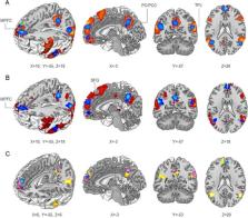
          The ability to understand mental states of others is referred to as mentalizing and enabled by our Theory of Mind. This social skill relies on brain regions comprising the mentalizing network as robustly observed in adults but also in a growing number of developmental studies. We summarized and compared neuroimaging evidence in children/adolescents and adults during mentalizing using coordinate-based activation likelihood estimation meta-analyses to inform about brain regions consistently or differentially engaged across age categories. Adults ( N = 5286) recruited medial prefrontal and middle/inferior frontal cortices, precuneus, temporoparietal junction and middle temporal gyri during mentalizing, which were functionally connected to bilateral inferior/superior parietal lobule and thalamus/striatum. Conjunction and contrast analyses revealed that children and adolescents ( N = 479) recruit similar but fewer regions within core mentalizing regions. Subgroup analyses revealed an early continuous engagement of middle medial prefrontal cortex, precuneus and right temporoparietal junction in younger children (8–11 years) and adolescents (12–18 years). Adolescents additionally recruited the left temporoparietal junction and middle/inferior temporal cortex. Overall, the observed engagement of the medial prefrontal cortex, precuneus and right temporoparietal junction during mentalizing across all ages reflects an early specialization of some key regions of the social brain.

          Related collections

          Most cited references118

          • Record: found
          • Abstract: found
          • Article: not found

          The adolescent brain and age-related behavioral manifestations.

          L Spear (2000)
          To successfully negotiate the developmental transition between youth and adulthood, adolescents must maneuver this often stressful period while acquiring skills necessary for independence. Certain behavioral features, including age-related increases in social behavior and risk-taking/novelty-seeking, are common among adolescents of diverse mammalian species and may aid in this process. Reduced positive incentive values from stimuli may lead adolescents to pursue new appetitive reinforcers through drug use and other risk-taking behaviors, with their relative insensitivity to drugs supporting comparatively greater per occasion use. Pubertal increases in gonadal hormones are a hallmark of adolescence, although there is little evidence for a simple association of these hormones with behavioral change during adolescence. Prominent developmental transformations are seen in prefrontal cortex and limbic brain regions of adolescents across a variety of species, alterations that include an apparent shift in the balance between mesocortical and mesolimbic dopamine systems. Developmental changes in these stressor-sensitive regions, which are critical for attributing incentive salience to drugs and other stimuli, likely contribute to the unique characteristics of adolescence.
            Bookmark
            • Record: found
            • Abstract: found
            • Article: not found

            The precuneus: a review of its functional anatomy and behavioural correlates.

            Functional neuroimaging studies have started unravelling unexpected functional attributes for the posteromedial portion of the parietal lobe, the precuneus. This cortical area has traditionally received little attention, mainly because of its hidden location and the virtual absence of focal lesion studies. However, recent functional imaging findings in healthy subjects suggest a central role for the precuneus in a wide spectrum of highly integrated tasks, including visuo-spatial imagery, episodic memory retrieval and self-processing operations, namely first-person perspective taking and an experience of agency. Furthermore, precuneus and surrounding posteromedial areas are amongst the brain structures displaying the highest resting metabolic rates (hot spots) and are characterized by transient decreases in the tonic activity during engagement in non-self-referential goal-directed actions (default mode of brain function). Therefore, it has recently been proposed that precuneus is involved in the interwoven network of the neural correlates of self-consciousness, engaged in self-related mental representations during rest. This hypothesis is consistent with the selective hypometabolism in the posteromedial cortex reported in a wide range of altered conscious states, such as sleep, drug-induced anaesthesia and vegetative states. This review summarizes the current knowledge about the macroscopic and microscopic anatomy of precuneus, together with its wide-spread connectivity with both cortical and subcortical structures, as shown by connectional and neurophysiological findings in non-human primates, and links these notions with the multifaceted spectrum of its behavioural correlates. By means of a critical analysis of precuneus activation patterns in response to different mental tasks, this paper provides a useful conceptual framework for matching the functional imaging findings with the specific role(s) played by this structure in the higher-order cognitive functions in which it has been implicated. Specifically, activation patterns appear to converge with anatomical and connectivity data in providing preliminary evidence for a functional subdivision within the precuneus into an anterior region, involved in self-centred mental imagery strategies, and a posterior region, subserving successful episodic memory retrieval.
              Bookmark
              • Record: found
              • Abstract: found
              • Article: not found

              Dynamic mapping of human cortical development during childhood through early adulthood.

              We report the dynamic anatomical sequence of human cortical gray matter development between the age of 4-21 years using quantitative four-dimensional maps and time-lapse sequences. Thirteen healthy children for whom anatomic brain MRI scans were obtained every 2 years, for 8-10 years, were studied. By using models of the cortical surface and sulcal landmarks and a statistical model for gray matter density, human cortical development could be visualized across the age range in a spatiotemporally detailed time-lapse sequence. The resulting time-lapse "movies" reveal that (i) higher-order association cortices mature only after lower-order somatosensory and visual cortices, the functions of which they integrate, are developed, and (ii) phylogenetically older brain areas mature earlier than newer ones. Direct comparison with normal cortical development may help understanding of some neurodevelopmental disorders such as childhood-onset schizophrenia or autism.
                Bookmark

                Author and article information

                Contributors
                Journal
                Soc Cogn Affect Neurosci
                Soc Cogn Affect Neurosci
                scan
                Social Cognitive and Affective Neuroscience
                Oxford University Press (UK )
                1749-5016
                1749-5024
                April 2022
                21 September 2021
                21 September 2021
                : 17
                : 4
                : 351-366
                Affiliations
                departmentJacobs Center for Productive Youth Development, University of Zurich , Zurich 8050, Switzerland
                departmentDepartment of Child and Adolescent Psychiatry, University of Basel, Psychiatric University Hospital , Basel 4002, Switzerland
                departmentJacobs Center for Productive Youth Development, University of Zurich , Zurich 8050, Switzerland
                departmentDepartment of Child and Adolescent Psychiatry, University of Basel, Psychiatric University Hospital , Basel 4002, Switzerland
                departmentDepartment of Child and Adolescent Psychiatry, University of Basel, Psychiatric University Hospital , Basel 4002, Switzerland
                departmentInstitute of Systems Neuroscience, Medical Faculty, Heinrich Heine University Düsseldorf , Düsseldorf 40225, Germany
                departmentBrain & Behaviour (INM-7), Research Centre Jülich, Institute of Neuroscience and Medicine , Jülich 52425, Germany
                departmentJacobs Center for Productive Youth Development, University of Zurich , Zurich 8050, Switzerland
                departmentDepartment of Child and Adolescent Psychiatry, University of Basel, Psychiatric University Hospital , Basel 4002, Switzerland
                departmentNeuroscience Center Zurich, University of Zurich and ETH Zurich , Zurich 8057, Switzerland
                Author notes
                Correspondence should be addressed to Nora M. Raschle, Jacobs Center for Productive Youth Development, University of Zurich, Andreasstrasse 15, Zürich 8050, Switzerland. E-mail: nora.raschle@ 123456jacobscenter.uzh.ch .
                Author information
                https://orcid.org/0000-0002-7649-8699
                https://orcid.org/0000-0002-3160-5999
                Article
                nsab105
                10.1093/scan/nsab105
                8972312
                34545389
                568ddb4d-b8a3-4cf2-9c31-ebd6989807d3
                © The Author(s) 2021. Published by Oxford University Press.

                This is an Open Access article distributed under the terms of the Creative Commons Attribution-NonCommercial License ( https://creativecommons.org/licenses/by-nc/4.0/), which permits non-commercial re-use, distribution, and reproduction in any medium, provided the original work is properly cited. For commercial re-use, please contact journals.permissions@oup.com

                History
                : 01 July 2020
                : 06 July 2021
                : 19 September 2021
                : 27 August 2021
                : 24 September 2021
                Page count
                Pages: 16
                Funding
                Funded by: Jacobs Foundation, DOI 10.13039/501100003986;
                Award ID: Early Career Research Grant [grant number 20162017
                Funded by: Universität Basel, DOI 10.13039/100008375;
                Award ID: Early career research grant
                Categories
                Original Manuscript
                AcademicSubjects/SCI01880

                Neurosciences
                mentalizing,functional neuroimaging,development,children,theory of mind,activation likelihood estimation

                Comments

                Comment on this article