- Record: found
- Abstract: found
- Article: not found
A Direct Comparison of Enhanced Saliva to Nasopharyngeal Swab for the Detection of SARS-CoV-2 in Symptomatic Patients
Read this article at

Abstract
The ongoing coronavirus disease 2019 (COVID-19) pandemic has resulted in shortages of nasopharyngeal swabs (NPS) and viral transport media, necessitating the search for alternate diagnostic specimens, such as saliva. We directly compared matched saliva and NPS specimens from symptomatic patients suspected of having COVID-19. An enhanced saliva specimen (i.e., strong sniff, elicited cough, and collection of saliva/secretions) was collected without transport medium prior to collection of NPS from 224 patients with symptoms deemed consistent with COVID-19.
ABSTRACT
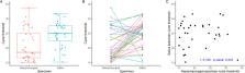
The ongoing coronavirus disease 2019 (COVID-19) pandemic has resulted in shortages of nasopharyngeal swabs (NPS) and viral transport media, necessitating the search for alternate diagnostic specimens, such as saliva. We directly compared matched saliva and NPS specimens from symptomatic patients suspected of having COVID-19. An enhanced saliva specimen (i.e., strong sniff, elicited cough, and collection of saliva/secretions) was collected without transport medium prior to collection of NPS from 224 patients with symptoms deemed consistent with COVID-19. Both specimens were tested with the CDC 2019 nCoV real-time RT-PCR diagnostic panel (4 February 2020 version), with the NPS result used as the reference standard. For the 216 patients included in the final analysis, there was 100% positive agreement (38/38 positive specimens) and 99.4% negative agreement (177/178 negative specimens). The one discrepant specimen had the presence of severe acute respiratory syndrome coronavirus 2 (SARS-CoV-2) confirmed in the saliva specimen using an alternate FDA EUA assay. The overall mean difference in cycle threshold ( C T ) values for the positive NPS and saliva specimens was −3.61 (95% confidence interval [CI], −5.78 to −1.44; P = 0.002). An enhanced saliva specimen performed as well as NPS for the qualitative detection of SARS-CoV-2 in symptomatic patients, although the overall mean viral load in saliva was lower.
Related collections
Most cited references20
- Record: found
- Abstract: found
- Article: not found
SARS-CoV-2 Viral Load in Upper Respiratory Specimens of Infected Patients
- Record: found
- Abstract: found
- Article: not found
Temporal profiles of viral load in posterior oropharyngeal saliva samples and serum antibody responses during infection by SARS-CoV-2: an observational cohort study
- Record: found
- Abstract: found
- Article: not found