8
views
0
recommends
+1 Recommend
0 collections
    0
    shares
      • Record: found
      • Abstract: found
      • Article: found
      Is Open Access

      Alteration of Vascular Responsiveness to Uridine Adenosine Tetraphosphate in Aortas Isolated from Male Diabetic Otsuka Long-Evans Tokushima Fatty Rats: The Involvement of Prostanoids

      research-article

      Read this article at

      Bookmark
          There is no author summary for this article yet. Authors can add summaries to their articles on ScienceOpen to make them more accessible to a non-specialist audience.

          Abstract

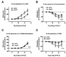
          We investigated whether responsiveness to dinucleotide uridine adenosine tetraphosphate (Up 4A) was altered in aortas from type 2 diabetic Otsuka Long-Evans Tokushima Fatty (OLETF) rats compared with those from age-matched control Long-Evans Tokushima Otsuka (LETO) rats at the chronic stage of disease. In OLETF aortas, we observed the following: (1) Up 4A-induced contractions were lower than those in the LETO aortas under basal conditions, (2) slight relaxation occurred due to Up 4A, but this was not observed in phenylephrine-precontracted LETO aortas, (3) acetylcholine-induced relaxation was reduced (vs. LETO), and (4) prostanoid release (prostaglandin (PG)F , thromboxane (Tx)A 2 metabolite, and PGE 2) due to Up 4A was decreased (vs. LETO). Endothelial denudation suppressed Up 4A-induced contractions in the LETO group, but increased the contractions in the OLETF group. Under nitric oxide synthase (NOS) inhibition, Up 4A induced contractions in phenylephrine-precontracted aortas; this effect was greater in the LETO group (vs. the OLETF group). The relaxation response induced by Up 4A was unmasked by cyclooxygenase inhibitors, especially in the LETO group, but this effect was abolished by NOS inhibition. These results suggest that the relaxant component of the Up 4A-mediated response was masked by prostanoids in the LETO aortas and that the LETO and OLETF rats presented different contributions of the endothelium to the response.

          Related collections

          Most cited references64

          • Record: found
          • Abstract: found
          • Article: not found

          Spontaneous long-term hyperglycemic rat with diabetic complications. Otsuka Long-Evans Tokushima Fatty (OLETF) strain.

          A spontaneously diabetic rat with polyuria, polydipsia, and mild obesity was discovered in 1984 in an outbred colony of Long-Evans rats, which had been purchased from Charles River Canada (St. Constant, Quebec, Canada) in 1982. A strain of rats developed from this rat by selective breeding has since been maintained at the Tokushima Research Institute (Otsuka Pharmaceutical, Tokushima, Japan) and named OLETF. The characteristic features of OLETF rats are 1) late onset of hyperglycemia (after 18 wk of age); 2) a chronic course of disease; 3) mild obesity; 4) inheritance by males; 5) hyperplastic foci of pancreatic islets; and 6) renal complication (nodular lesions). Histologically, the changes of pancreatic islets can be classified into three stages: 1) an early stage (6-20 wk of age) of cellular infiltration and degeneration; 2) a hyperplastic stage (20-40 wk of age); and 3) a final stage (at > 40 wk of age). These clinical and pathological features of disease in OLETF rats resemble those of human NIDDM.
            Bookmark
            • Record: found
            • Abstract: found
            • Article: not found

            Thirty Years of Saying NO: Sources, Fate, Actions, and Misfortunes of the Endothelium-Derived Vasodilator Mediator.

            Endothelial cells control vascular tone by releasing nitric oxide (NO) produced by endothelial NO synthase. The activity of endothelial NO synthase is modulated by the calcium concentration and by post-translational modifications (eg, phosphorylation). When NO reaches vascular smooth muscle, soluble guanylyl cyclase is its primary target producing cGMP. NO production is stimulated by circulating substances (eg, catecholamines), platelet products (eg, serotonin), autacoids formed in (eg, bradykinin) or near (eg, adiponectin) the vascular wall and physical factors (eg, shear stress). NO dysfunction can be caused, alone or in combination, by abnormal coupling of endothelial cell membrane receptors, insufficient supply of substrate (l-arginine) or cofactors (tetrahydrobiopterin), endogenous inhibitors (asymmetrical dimethyl arginine), reduced expression/presence/dimerization of endothelial NO synthase, inhibition of its enzymatic activity, accelerated disposition of NO by reactive oxygen species and abnormal responses (eg, biased soluble guanylyl cyclase activity producing cyclic inosine monophosphate) of the vascular smooth muscle. Major culprits causing endothelial dysfunction, irrespective of the underlying pathological process (aging, obesity, diabetes mellitus, and hypertension), include stimulation of mineralocorticoid receptors, activation of endothelial Rho-kinase, augmented presence of asymmetrical dimethyl arginine, and exaggerated oxidative stress. Genetic and pharmacological interventions improve dysfunctional NO-mediated vasodilatations if protecting the supply of substrate and cofactors for endothelial NO synthase, preserving the presence and activity of the enzyme and reducing reactive oxygen species generation. Common achievers of such improvement include maintained levels of estrogens and increased production of adiponectin and induction of silent mating-type information regulation 2 homologue 1. Obviously, endothelium-dependent relaxations are not the only beneficial action of NO in the vascular wall. Thus, reduced NO-mediated responses precede and initiate the atherosclerotic process.
              Bookmark
              • Record: found
              • Abstract: found
              • Article: not found

              Purinergic Signaling in the Cardiovascular System.

              There is nervous control of the heart by ATP as a cotransmitter in sympathetic, parasympathetic, and sensory-motor nerves, as well as in intracardiac neurons. Centers in the brain control heart activities and vagal cardiovascular reflexes involve purines. Adenine nucleotides and nucleosides act on purinoceptors on cardiomyocytes, AV and SA nodes, cardiac fibroblasts, and coronary blood vessels. Vascular tone is controlled by a dual mechanism. ATP, released from perivascular sympathetic nerves, causes vasoconstriction largely via P2X1 receptors. Endothelial cells release ATP in response to changes in blood flow (via shear stress) or hypoxia, to act on P2 receptors on endothelial cells to produce nitric oxide, endothelium-derived hyperpolarizing factor, or prostaglandins to cause vasodilation. ATP is also released from sensory-motor nerves during antidromic reflex activity, to produce relaxation of some blood vessels. Purinergic signaling is involved in the physiology of erythrocytes, platelets, and leukocytes. ATP is released from erythrocytes and platelets, and purinoceptors and ectonucleotidases are expressed by these cells. P1, P2Y1, P2Y12, and P2X1 receptors are expressed on platelets, which mediate platelet aggregation and shape change. Long-term (trophic) actions of purine and pyrimidine nucleosides and nucleotides promote migration and proliferation of vascular smooth muscle and endothelial cells via P1 and P2Y receptors during angiogenesis, vessel remodeling during restenosis after angioplasty and atherosclerosis. The involvement of purinergic signaling in cardiovascular pathophysiology and its therapeutic potential are discussed, including heart failure, infarction, arrhythmias, syncope, cardiomyopathy, angina, heart transplantation and coronary bypass grafts, coronary artery disease, diabetic cardiomyopathy, hypertension, ischemia, thrombosis, diabetes mellitus, and migraine.
                Bookmark

                Author and article information

                Journal
                Int J Mol Sci
                Int J Mol Sci
                ijms
                International Journal of Molecular Sciences
                MDPI
                1422-0067
                09 November 2017
                November 2017
                : 18
                : 11
                : 2378
                Affiliations
                Department of Physiology and Morphology, Institute of Medicinal Chemistry, Hoshi University, Shinagawa-ku, Tokyo 142-8501, Japan; sho.kobayan.1213@ 123456gmail.com (S.K.); redevil1126@ 123456gmail.com (M.A.); maika.iguchi@ 123456gmail.com (M.I.); k955tkyng@ 123456gmail.com (K.T.); cozymihomiho.0210@ 123456gmail.com (M.K.); k-taguchi@ 123456hoshi.ac.jp (K.T.)
                Author notes
                [* ]Correspondence: t-matsu@ 123456hoshi.ac.jp (T.M.); tkoba@ 123456hoshi.ac.jp (T.K.); Tel./Fax: +81-3-5498-5726 (T.M.); +81-3-5498-5849 (T.K.)
                Author information
                https://orcid.org/0000-0001-9177-8384
                Article
                ijms-18-02378
                10.3390/ijms18112378
                5713347
                29120387
                57212759-f65a-4764-955b-7d64e5df0be3
                © 2017 by the authors.

                Licensee MDPI, Basel, Switzerland. This article is an open access article distributed under the terms and conditions of the Creative Commons Attribution (CC BY) license ( http://creativecommons.org/licenses/by/4.0/).

                History
                : 27 October 2017
                : 08 November 2017
                Categories
                Article

                Molecular biology
                aorta,contraction,prostanoid,relaxation,type 2 diabetes,up4a
                Molecular biology
                aorta, contraction, prostanoid, relaxation, type 2 diabetes, up4a

                Comments

                Comment on this article