13
views
0
recommends
+1 Recommend
0 collections
    0
    shares
      • Record: found
      • Abstract: found
      • Article: found
      Is Open Access

      Endoscopic resection of acetabular screw tip to decompress sciatic nerve following total hip arthroplasty

      case-report

      Read this article at

      Bookmark
          There is no author summary for this article yet. Authors can add summaries to their articles on ScienceOpen to make them more accessible to a non-specialist audience.

          Abstract

          Background

          Sciatic nerve injuries following total hip arthroplasty are disabling complications. Although degrees of injury are variable from neuropraxia to neurotmesis, mechanical irritation of sciatic nerve might be occurred by protruding hardware. This case shows endoscopic decompression for protruded acetabular screw irritating sciatic nerve, the techniques described herein may permit broader arthroscopic/endoscopic applications for management of complications after reconstructive hip surgery.

          Case presentation

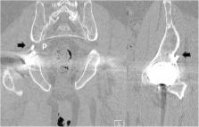
          An 80-year-old man complained of severe pain and paresthesias following acetabular component revision surgery. Physical findings included right buttock pain with radiating pain to lower extremity. Radiographs and computed tomography imaging showed that the sharp end of protruded screw invaded greater sciatic foramen anterior to posterior and distal to proximal direction at sciatic notch level. A protruding tip of the acetabular screw at the sciatic notch was decompressed by use of techniques gained from experience performing endoscopic sciatic nerve decompression. The pre-operative pain and paresthesias resolved post-operatively after recovering from anesthesia.

          Conclusions

          This case report describes the first documented endoscopic resection of the tip of the acetabular screw irritating sciatic nerve after total hip arthroplasty. If endoscopic resection of an offending acetabular screw can be performed in a safe and minimally invasive manner, one can envision a future expansion of the role of hip arthroscopic surgery in several complications management after total hip arthroplasty.

          Electronic supplementary material

          The online version of this article (10.1186/s12891-018-2091-x) contains supplementary material, which is available to authorized users.

          Related collections

          Most cited references7

          • Record: found
          • Abstract: found
          • Article: not found

          Acetabular anatomy and the transacetabular fixation of screws in total hip arthroplasty.

          An anatomical and radiographic study was undertaken to determine the safest zones in the acetabulum for the transacetabular placement of screws during uncemented acetabular arthroplasty. To avoid injury to intrapelvic structures, which are not visible to the surgeon during placement of the screws, cadavera were studied to define the location of these structures with respect to fixed points of reference within the acetabulum. Four clinically useful acetabular quadrants were delineated. The quadrants are formed by drawing a line from the anterior superior iliac spine through the center of the acetabulum to the posterior fovea, forming acetabular halves. A second line is then drawn perpendicular to the first at the mid-point of the acetabulum, forming four quadrants. The posterior superior and posterior inferior acetabular quadrants contain the best available bone stock and are relatively safe for the transacetabular placement of screws. The anterior superior and anterior inferior quandrants should be avoided whenever possible, because screws placed improperly in these quadrants may endanger the external iliac artery and vein, as well as the obturator nerve, artery, and vein. The acetabular-quadrant system provides the surgeon with a simple intraoperative guide to the safe transacetabular placement of screws during primary and revision acetabular arthroplasty.
            Bookmark
            • Record: found
            • Abstract: found
            • Article: not found

            Update on nerve palsy associated with total hip replacement.

            Nerve palsy is an uncommon but acknowledged complication of total hip replacement. The overall prevalence is approximately 1%. The sciatic nerve, or the peroneal division of the sciatic nerve, is involved in nearly 80% of cases. The risk of nerve palsy in association with total hip replacement is increased for female compared with male patients, with a diagnosis of developmental dysplasia, and with patients undergoing revision surgery. In the majority of cases, the origin of the palsy is unknown. Because peripheral nerves are sensitive to compression, unrecognized compression may play a role in these cases. The prognosis for neurologic recovery is related to the degree of nerve damage. Complete, or essentially complete, recovery occurs in approximately 41% and another 44% have only a mild deficit. Approximately 15% have a poor outcome characterized by weakness that limits ambulation and/or persistent dysesthesia. Patients with some motor function immediately after the operation and those who recover some motor function within approximately 2 weeks of surgery have a good prognosis for recovery. In general, recovery of femoral nerve palsies is more predictable than that of sciatic palsies.
              Bookmark
              • Record: found
              • Abstract: found
              • Article: not found

              Neurologic injuries after total hip arthroplasty.

              Neurologic injuries are a potentially devastating complication of total hip arthroplasty (THA). Review of the literature reveals that these injuries are uncommon. The reported incidence ranges from 0.08% to 7.6%. The incidence in primary THA ranges from 0.09% to 3.7% and in revision THA from 0% to 7.6%. Reported etiologies include intraoperative direct nerve injury, significant leg lengthening, improper retractor placement, cement extravasation, cement-related thermal damage, patient positioning, manipulation, and postoperative hematoma. Risk factors include developmental dysplasia of the hip, the female sex, posttraumatic arthritis, and revision surgery. However, no single risk factor has been consistently reported to be significant, and many patients with no known risk factors incur neurologic injuries.
                Bookmark

                Author and article information

                Contributors
                82-63-250-2798 , sunjungyoonkos@gmail.com
                20355@cuh.co.kr
                saltandlight777@hotmail.com
                why76@jbnu.ac.kr
                Journal
                BMC Musculoskelet Disord
                BMC Musculoskelet Disord
                BMC Musculoskeletal Disorders
                BioMed Central (London )
                1471-2474
                4 June 2018
                4 June 2018
                2018
                : 19
                : 184
                Affiliations
                [1 ]ISNI 0000 0004 0647 1516, GRID grid.411551.5, Department of Orthopedic Surgery, , Research Institute of clinical medicine of Chonbuk National University, Biomedical Research Institute of Chonbuk National University Hospital, ; 54907 Gunji-Ro 20, Dukjin-Gu, Chonbuk, Jeonju, South Korea
                [2 ]DISC Sports and Spine Center, Marina del Rey, CA USA
                [3 ]ISNI 0000 0004 0470 4320, GRID grid.411545.0, Department of Anatomy, , Medical School, Institute for Medical Sciences, Chonbuk National University, ; Jeonju, 561-180 South Korea
                Author information
                http://orcid.org/0000-0003-4943-534X
                Article
                2091
                10.1186/s12891-018-2091-x
                5987599
                29866097
                57997b27-50f8-47aa-97ab-b53dcc2dc3f0
                © The Author(s). 2018

                Open AccessThis article is distributed under the terms of the Creative Commons Attribution 4.0 International License ( http://creativecommons.org/licenses/by/4.0/), which permits unrestricted use, distribution, and reproduction in any medium, provided you give appropriate credit to the original author(s) and the source, provide a link to the Creative Commons license, and indicate if changes were made. The Creative Commons Public Domain Dedication waiver ( http://creativecommons.org/publicdomain/zero/1.0/) applies to the data made available in this article, unless otherwise stated.

                History
                : 30 January 2018
                : 15 May 2018
                Categories
                Case Report
                Custom metadata
                © The Author(s) 2018

                Orthopedics
                endoscopic sciatic nerve decompression,sciatic nerve neuropathy,acetabular dome screw,total hip arthroplasty

                Comments

                Comment on this article