18
views
0
recommends
+1 Recommend
0 collections
    0
    shares
      • Record: found
      • Abstract: found
      • Article: found
      Is Open Access

      Grass Carp Laboratory of Genetics and Physiology 2 Serves As a Negative Regulator in Retinoic Acid-Inducible Gene I- and Melanoma Differentiation-Associated Gene 5-Mediated Antiviral Signaling in Resting State and Early Stage of Grass Carp Reovirus Infection

      research-article

      Read this article at

      Bookmark
          There is no author summary for this article yet. Authors can add summaries to their articles on ScienceOpen to make them more accessible to a non-specialist audience.

          Abstract

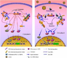
          Laboratory of genetics and physiology 2 (LGP2) is a key component of RIG-I-like receptors (RLRs). However, the lack of the caspase recruitment domains (CARDs) results in its controversial functional performance as a negative or positive regulator in antiviral responses. Especially, no sufficient evidence uncovers the functional mechanisms of LGP2 in RLR signaling pathways in teleost. Here, negative regulation mechanism of LGP2 in certain situations in retinoic acid-inducible gene I (RIG-I) and melanoma differentiation-associated gene 5 (MDA5)-mediated antiviral responses was identified in Ctenopharyngodon idella kidney cells. LGP2 overexpression inhibits synthesis and phosphorylation of interferon regulatory factor 3/7 (IRF3/7), and mRNA levels and promoter activities of IFNs and NF-κBs in resting state and early phase of grass carp reovirus (GCRV) infection. Knockdown of LGP2 obtains opposite effects. Luciferase report assay indicates that LGP2 works at the upstream of RIG-I and MDA5. LGP2 binds to RIG-I and MDA5 with diverse domain preference and which is independent of GCRV infection. Furthermore, LGP2 restrains K63-linked ubiquitination of RIG-I and MDA5 in various degrees. These differences result in disparate repressive mechanisms of LGP2 to RIG-I- and MDA5-mediated signal activations of IFN-β promoter stimulator 1 and mediator of IRF3 activation. Interestingly, LGP2 also inhibits K48-linked RIG-I and MDA5 ubiquitination to suppress proteins degradation, which guarantees the basal protein levels for subsequently rapid signal activation. All these results reveal a mechanism that LGP2 functions as a suppressor in RLR signaling pathways to maintain cellular homeostasis in resting state and early phase during GCRV infection.

          Related collections

          Most cited references50

          • Record: found
          • Abstract: found
          • Article: not found

          Structural basis for the activation of innate immune pattern-recognition receptor RIG-I by viral RNA.

          RIG-I is a key innate immune pattern-recognition receptor that triggers interferon expression upon detection of intracellular 5'triphosphate double-stranded RNA (5'ppp-dsRNA) of viral origin. RIG-I comprises N-terminal caspase activation and recruitment domains (CARDs), a DECH helicase, and a C-terminal domain (CTD). We present crystal structures of the ligand-free, autorepressed, and RNA-bound, activated states of RIG-I. Inactive RIG-I has an open conformation with the CARDs sequestered by a helical domain inserted between the two helicase moieties. ATP and dsRNA binding induce a major rearrangement to a closed conformation in which the helicase and CTD bind the blunt end 5'ppp-dsRNA with perfect complementarity but incompatibly with continued CARD binding. We propose that after initial binding of 5'ppp-dsRNA to the flexibly linked CTD, co-operative tight binding of ATP and RNA to the helicase domain liberates the CARDs for downstream signaling. These findings significantly advance our molecular understanding of the activation of innate immune signaling helicases. Copyright © 2011 Elsevier Inc. All rights reserved.
            Bookmark
            • Record: found
            • Abstract: found
            • Article: not found

            Regulation of innate antiviral defenses through a shared repressor domain in RIG-I and LGP2.

            RIG-I is an RNA helicase containing caspase activation and recruitment domains (CARDs). RNA binding and signaling by RIG-I are implicated in pathogen recognition and triggering of IFN-alpha/beta immune defenses that impact cell permissiveness for hepatitis C virus (HCV). Here we evaluated the processes that control RIG-I signaling. RNA binding studies and analysis of cells lacking RIG-I, or the related MDA5 protein, demonstrated that RIG-I, but not MDA5, efficiently binds to secondary structured HCV RNA to confer induction of IFN-beta expression. We also found that LGP2, a helicase related to RIG-I and MDA5 but lacking CARDs and functioning as a negative regulator of host defense, binds HCV RNA. In resting cells, RIG-I is maintained as a monomer in an autoinhibited state, but during virus infection and RNA binding it undergoes a conformation shift that promotes self-association and CARD interactions with the IPS-1 adaptor protein to signal IFN regulatory factor 3- and NF-kappaB-responsive genes. This reaction is governed by an internal repressor domain (RD) that controls RIG-I multimerization and IPS-1 interaction. Deletion of the RIG-I RD resulted in constitutive signaling to the IFN-beta promoter, whereas RD expression alone prevented signaling and increased cellular permissiveness to HCV. We identified an analogous RD within LGP2 that interacts in trans with RIG-I to ablate self-association and signaling. Thus, RIG-I is a cytoplasmic sensor of HCV and is governed by RD interactions that are shared with LGP2 as an on/off switch controlling innate defenses. Modulation of RIG-I/LGP2 interaction dynamics may have therapeutic implications for immune regulation.
              Bookmark
              • Record: found
              • Abstract: found
              • Article: not found

              Reconstitution of the RIG-I pathway reveals a signaling role of unanchored polyubiquitin chains in innate immunity.

              RIG-I detects invading viral RNA and activates the transcription factors NF-kappaB and IRF3 through the mitochondrial protein MAVS. Here we show that RNA bearing 5'-triphosphate strongly activates the RIG-I-IRF3 signaling cascade in a reconstituted system composed of RIG-I, mitochondria, and cytosol. Activation of RIG-I requires not only RNA but also polyubiquitin chains linked through lysine 63 (K63) of ubiquitin. RIG-I binds specifically to K63-polyubiquitin chains through its tandem CARD domains in a manner that depends on RNA and ATP. Mutations in the CARD domains that abrogate ubiquitin binding also impair RIG-I activation. Remarkably, unanchored K63-ubiquitin chains, which are not conjugated to any target protein, potently activate RIG-I. These ubiquitin chains function as an endogenous ligand of RIG-I in human cells. Our results delineate the mechanism of RIG-I activation, identify CARD domains as a ubiquitin sensor, and demonstrate that unanchored K63-polyubiquitin chains are signaling molecules in antiviral innate immunity. Copyright 2010 Elsevier Inc. All rights reserved.
                Bookmark

                Author and article information

                Contributors
                Journal
                Front Immunol
                Front Immunol
                Front. Immunol.
                Frontiers in Immunology
                Frontiers Media S.A.
                1664-3224
                27 March 2017
                2017
                : 8
                : 352
                Affiliations
                [1] 1College of Fisheries, Huazhong Agricultural University , Wuhan, China
                [2] 2College of Veterinary Medicine, Huazhong Agricultural University , Wuhan, China
                Author notes

                Edited by: Claudia U. Duerr, McGill University, Canada

                Reviewed by: Alan G. Goodman, Washington State University, USA; Juan Pablo De Rivero Vaccari, University of Miami, USA

                *Correspondence: Jianguo Su, sujianguo@ 123456mail.hzau.edu.cn

                Specialty section: This article was submitted to Molecular Innate Immunity, a section of the journal Frontiers in Immunology

                Article
                10.3389/fimmu.2017.00352
                5366347
                28396670
                5a73d01e-2ef7-4ba9-a82f-f01d10d4606c
                Copyright © 2017 Rao, Wan, Yang and Su.

                This is an open-access article distributed under the terms of the Creative Commons Attribution License (CC BY). The use, distribution or reproduction in other forums is permitted, provided the original author(s) or licensor are credited and that the original publication in this journal is cited, in accordance with accepted academic practice. No use, distribution or reproduction is permitted which does not comply with these terms.

                History
                : 20 January 2017
                : 13 March 2017
                Page count
                Figures: 9, Tables: 0, Equations: 0, References: 57, Pages: 15, Words: 10011
                Funding
                Funded by: National Natural Science Foundation of China 10.13039/501100001809
                Award ID: 31572648
                Categories
                Immunology
                Original Research

                Immunology
                laboratory of genetics and physiology 2,innate immunity,grass carp (ctenopharyngodon idella),grass carp reovirus,rlrs,interferon regulatory factor 3,irf7

                Comments

                Comment on this article