3
views
0
recommends
+1 Recommend
0 collections
    0
    shares
      • Record: found
      • Abstract: found
      • Article: found
      Is Open Access

      Increased Beat-to-Beat Variability of T-Wave Heterogeneity Measured From Standard 12-Lead Electrocardiogram Is Associated With Sudden Cardiac Death: A Case–Control Study

      research-article

      Read this article at

      Bookmark
          There is no author summary for this article yet. Authors can add summaries to their articles on ScienceOpen to make them more accessible to a non-specialist audience.

          Abstract

          Introduction

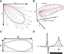
          The prognostic significance of beat-to-beat variability of spatial heterogeneity of repolarization measured from standard 12-lead ECG is not well-understood.

          Methods

          We measured the short-term variability of repolarization parameters, such as T-wave heterogeneity in leads V4–V6 (TWH) and QT interval (QT), from five consecutive beats of previously recorded standard 12-lead ECG in 200 victims of unexpected sudden cardiac death (SCD) confirmed to be due to complicated atherosclerotic coronary artery disease (CAD) in medico-legal autopsy and 200 age- and sex-matched controls with angiographically confirmed CAD. The short-term variability of repolarization heterogeneity was defined as the standard deviation (SD) of the measured repolarization parameters. All ECGs were in sinus rhythm, and no premature ventricular contractions were included in the measured segment.

          Results

          TWH-SD and QT-SD were significantly higher in SCD victims than in subjects with CAD (6.9 ± 5.6 μV vs. 3.8 ± 2.6 μV, p = 1.8E-11; 8.3 ± 13.1 ms vs. 3.8 ± 7.1 ms, p = 0.00003, respectively). After adjusting in the multivariate clinical model with factors, such as diabetes, RR interval, and beta blocker medication, TWH-SD and QT-SD retained their significant power in discriminating between the victims of SCD and the patients with CAD ( p = 0.00003, p = 0.006, respectively). TWH-SD outperformed QT-SD in identifying the SCD victims among the study subjects (area under the curve in the receiver operating characteristics curve 0.730 vs. 0.679, respectively).

          Conclusion

          Increased short-term variability of repolarization heterogeneity measured from standard 12-lead ECG is associated with SCD.

          Related collections

          Most cited references20

          • Record: found
          • Abstract: found
          • Article: not found

          QT interval prolongation predicts cardiovascular mortality in an apparently healthy population.

          In myocardial infarction patients, heart rate-adjusted QT interval (QTc), an electrocardiographic indicator of sympathetic balance, is prognostic for survival. In a 28-year follow-up, the association between QTc and all-cause, cardiovascular, and ischemic heart disease mortality was studied in a population of 3,091 apparently healthy Dutch civil servants and their spouses, aged 40-65 years, who participated in a medical examination during 1953-1954. Moderate (QTc, 420-440 msec) and extensive (QTc, more than 440 msec) QTc prolongations significantly predict all-cause mortality during the first 15 years among men (adjusted respective relative risks [RRs], 1.5 and 1.7) and among women (RRs, 1.7 and 1.6). In men, cardiovascular (RRs, 1.6 and 1.8) and ischemic heart disease mortality (RRs, 1.8 and 2.1) mainly account for this association. In women, the association cannot be attributed specifically to cardiovascular and ischemic heart disease mortality. RRs for a subpopulation without any sign of heart disease at baseline are similar. The same is observed for QTc prolongation after light exercise, although in this situation most associations are not statistically significant, probably because of smaller numbers in the QTc prolongation categories. Our results suggest that QTc contributes independently to cardiovascular risk. If autonomic imbalance is an important mechanism, it might be speculated that changes in life-style (e.g., with regard to physical exercise and smoking) may have a preventive impact.
            Bookmark
            • Record: found
            • Abstract: found
            • Article: not found

            QT interval prolongation as predictor of sudden death in patients with myocardial infarction.

            Fifty-five patients with recent myocardial infarction and 55 healthy controls, matched for age, sex, race, height, weight, education and job, had an electrocardiogram taken every two months for seven years. Twenty-eight patients and one control had a sudden cardiac death. The QTc (mean of all values recorded) was found prolonged in one control (2%), five of 27 surviving patients (18%) and in 16 of 28 patients who had sudden death (57%). The difference between surviving and sudden death patients is significant (P less than 0.01). It is interesting that the only control with a long QT was the one when died suddenly of myocardial infarction. Among patients with previous myocardial infarction a prolonged QTc constitutes a 2.16 times greater risk for sudden death. We conclude that a constant prolongation of QTc in patients with myocardial infarction may help, with other risk factors, in defining a subgroup at higher risk for sudden death.
              Bookmark
              • Record: found
              • Abstract: found
              • Article: not found

              QT interval variability and spontaneous ventricular tachycardia or fibrillation in the Multicenter Automatic Defibrillator Implantation Trial (MADIT) II patients.

              This study aimed to determine whether increased QT interval variability is associated with an increased risk for ventricular tachycardia (VT) or ventricular fibrillation (VF), documented by interrogation of the implantable cardioverter-defibrillator (ICD), in patients enrolled in the Multicenter Automatic Defibrillator Implantation Trial (MADIT) II. Unstable repolarization has been proposed as a risk factor for re-entrant arrhythmias, but confirmatory data from clinical trials are lacking. The QT variability was assessed in 10-min, resting high-resolution electrocardiogram recordings at study entry using a semiautomated algorithm that measured beat-to-beat QT duration in 817 MADIT II patients. The incidence of VT/VF requiring device therapy was determined by ICD interrogation. Median normalized QT variability (QTVN) was 0.179 and 0.125, respectively, in patients with VT/VF versus those without VT/VF (p = 0.001); QTVI (QTVN adjusted for heart rate variance) also was significantly (p < 0.05) higher in VT/VF patients than in those without VT/VF. Either QTVN or QTVI was linked with a significantly higher probability of VT/VF: two-year risk of VT/VF from Kaplan-Meier curves was 40% in highest quartile versus 21% in lower quartiles for QTVN, and 37% versus 22% for QTVI (p < 0.05 for each). In multivariate Cox regression models adjusting for clinical covariates (race, New York Heart Association functional class, time after myocardial infarction), top-quartile QTVI and QTVN were independently associated with VT/VF (hazard ratio for QTVN 2.18, 95%confidence interval [CI] 1.34 to 3.55, p = 0.002; hazard ratio for QTVI 1.80, 95% CI 1.09 to 2.95, p = 0.021). In postinfarction patients with severe left ventricular dysfunction, increased QT variability, a marker of repolarization lability, is associated with an increased risk for VT/VF.
                Bookmark

                Author and article information

                Contributors
                Journal
                Front Physiol
                Front Physiol
                Front. Physiol.
                Frontiers in Physiology
                Frontiers Media S.A.
                1664-042X
                25 August 2020
                2020
                : 11
                : 1045
                Affiliations
                [1] 1Research Unit of Internal Medicine, Medical Research Center Oulu, Oulu University Hospital, University of Oulu , Oulu, Finland
                [2] 2Forensic Medicine Unit, Finnish Institute for Health and Welfare , Oulu, Finland
                [3] 3Research Unit of Internal Medicine, Department of Forensic Medicine, Medical Research Center Oulu, University of Oulu , Oulu, Finland
                Author notes

                Edited by: Marcel van der Heyden, University Medical Center Utrecht, Netherlands

                Reviewed by: Wojciech Zareba, University of Rochester, United States; Vijay S. Chauhan, Peter Munk Cardiac Centre, Canada; Juan Pablo Martínez, University of Zaragoza, Spain

                *Correspondence: Juha S. Perkiömäki, juha.perkiomaki@ 123456oulu.fi

                This article was submitted to Cardiac Electrophysiology, a section of the journal Frontiers in Physiology

                Article
                10.3389/fphys.2020.01045
                7477294
                5b8a0411-4039-4d5e-96e5-1562171ad48c
                Copyright © 2020 Hekkanen, Kenttä, Haukilahti, Rahola, Holmström, Vähätalo, Tulppo, Kiviniemi, Pakanen, Ukkola, Junttila, Huikuri and Perkiömäki.

                This is an open-access article distributed under the terms of the Creative Commons Attribution License (CC BY). The use, distribution or reproduction in other forums is permitted, provided the original author(s) and the copyright owner(s) are credited and that the original publication in this journal is cited, in accordance with accepted academic practice. No use, distribution or reproduction is permitted which does not comply with these terms.

                History
                : 09 June 2020
                : 29 July 2020
                Page count
                Figures: 2, Tables: 6, Equations: 0, References: 25, Pages: 9, Words: 0
                Categories
                Physiology
                Original Research

                Anatomy & Physiology
                electrocardiography,t-wave,t-wave morphology,repolarization,sudden cardiac death,ventricular arrhythmias

                Comments

                Comment on this article