23
views
0
recommends
+1 Recommend
0 collections
    0
    shares
      • Record: found
      • Abstract: found
      • Article: found
      Is Open Access

      Mast Cells Modulate Acute Toxoplasmosis in Murine Models

      research-article

      Read this article at
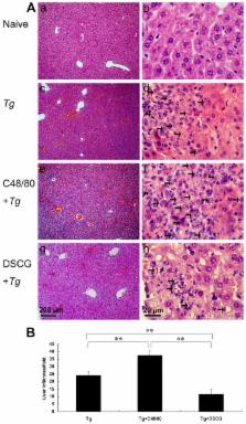
      Bookmark
          There is no author summary for this article yet. Authors can add summaries to their articles on ScienceOpen to make them more accessible to a non-specialist audience.

          Abstract

          The role of mast cells (MCs) in Toxoplasma gondii infection is poorly known. Kunming outbred mice were infected intraperitoneally with RH strain T. gondii, either treated with compound 48/80 (C48/80, MC activator) or disodium cromoglycate (DSCG, MC inhibitor). Compared with infected controls, infected mice treated with C48/80 exhibited significantly increased inflammation in the liver ( P < 0.01), spleen ( P < 0.05), and mesentery ( P < 0.05) tissues, higher parasite burden in the peritoneal lavage fluids ( P < 0.01), and increased levels of mRNA transcripts of T. gondii tachyzoite surface antigen 1 (SAG1) gene in the spleen and liver tissues ( P < 0.01), accompanied with significantly increased Th1 cytokine (IFN-γ, IL-12p40, and TNF-α) ( P < 0.01) and decreased IL-10 ( P < 0.01) mRNA expressions in the liver, and increased IFN-γ ( P < 0.01) and IL-12p40 ( P < 0.01) but decreased TNF-α ( P < 0.01) and IL-4 ( P < 0.01) in the spleens of infected mice treated with C48/80 at day 9-10 p.i. Whereas mice treated with DSCG had significantly decreased tissue lesions ( P < 0.01), lower parasite burden in the peritoneal lavage fluids ( P < 0.01) and decreased SAG1 expressions in the spleen and liver tissues ( P < 0.01), accompanied with significantly increased IFN-γ ( P < 0.01) and IL-12p40 ( P < 0.05) in the liver, and decreased IFN-γ ( P < 0.05) and TNF-α ( P < 0.01) in the spleens; IL-4 and IL-10 expressions in both the spleen and liver were significantly increased ( P < 0.01) in the infected mice treated with DSCG. These findings suggest that mediators associated with the MC activation may play an important role in modulating acute inflammatory pathogenesis and parasite clearance during T. gondii infection in this strain of mice. Thus, MC activation/inhibition mechanisms are potential novel targets for the prevention and control of T. gondii infection.

          Related collections

          Most cited references43

          • Record: found
          • Abstract: found
          • Article: not found

          Mast cells are essential intermediaries in regulatory T-cell tolerance.

          Contrary to the proinflammatory role of mast cells in allergic disorders, the results obtained in this study establish that mast cells are essential in CD4+CD25+Foxp3+ regulatory T (T(Reg))-cell-dependent peripheral tolerance. Here we confirm that tolerant allografts, which are sustained owing to the immunosuppressive effects of T(Reg) cells, acquire a unique genetic signature dominated by the expression of mast-cell-gene products. We also show that mast cells are crucial for allograft tolerance, through the inability to induce tolerance in mast-cell-deficient mice. High levels of interleukin (IL)-9--a mast cell growth and activation factor--are produced by activated T(Reg) cells, and IL-9 production seems important in mast cell recruitment to, and activation in, tolerant tissue. Our data indicate that IL-9 represents the functional link through which activated T(Reg) cells recruit and activate mast cells to mediate regional immune suppression, because neutralization of IL-9 greatly accelerates allograft rejection in tolerant mice. Finally, immunohistochemical analysis clearly demonstrates the existence of this novel T(Reg)-IL-9-mast cell relationship within tolerant allografts.
            Bookmark
            • Record: found
            • Abstract: found
            • Article: not found

            Mast cells.

            Mast cells are found resident in tissues throughout the body, particularly in association with structures such as blood vessels and nerves, and in proximity to surfaces that interface the external environment. Mast cells are bone marrow-derived and particularly depend upon stem cell factor for their survival. Mast cells express a variety of phenotypic features within tissues as determined by the local environment. Withdrawal of required growth factors results in mast cell apoptosis. Mast cells appear to be highly engineered cells with multiple critical biological functions. They may be activated by a number of stimuli that are both Fc epsilon RI dependent and Fc epsilon RI independent. Activation through various receptors leads to distinct signaling pathways. After activation, mast cells may immediately extrude granule-associated mediators and generate lipid-derived substances that induce immediate allergic inflammation. Mast cell activation may also be followed by the synthesis of chemokines and cytokines. Cytokine and chemokine secretion, which occurs hours later, may contribute to chronic inflammation. Biological functions of mast cells appear to include a role in innate immunity, involvement in host defense mechanisms against parasitic infestations, immunomodulation of the immune system, and tissue repair and angiogenesis.
              Bookmark
              • Record: found
              • Abstract: found
              • Article: not found

              Mast cell-derived interleukin 10 limits skin pathology in contact dermatitis and chronic irradiation with ultraviolet B.

              Allergic contact dermatitis, such as in response to poison ivy or poison oak, and chronic low-dose ultraviolet B irradiation can damage the skin. Mast cells produce proinflammatory mediators that are thought to exacerbate these prevalent acquired immune or innate responses. Here we found that, unexpectedly, mast cells substantially limited the pathology associated with these responses, including infiltrates of leukocytes, epidermal hyperplasia and epidermal necrosis. Production of interleukin 10 by mast cells contributed to the anti-inflammatory or immunosuppressive effects of mast cells in these conditions. Our findings identify a previously unrecognized function for mast cells and mast cell-derived interleukin 10 in limiting leukocyte infiltration, inflammation and tissue damage associated with immunological or innate responses that can injure the skin.
                Bookmark

                Author and article information

                Contributors
                Role: Editor
                Journal
                PLoS One
                PLoS ONE
                plos
                plosone
                PLoS ONE
                Public Library of Science (San Francisco, USA )
                1932-6203
                2013
                16 October 2013
                : 8
                : 10
                : e77327
                Affiliations
                [1 ]Department of Parasitology, Zhongshan School of Medicine, Sun Yat-sen University, Guangzhou, Guangdong, China
                [2 ]Key Laboratory of Tropical Disease Control (Sun Yat-sen University), Ministry of Education, Guangzhou, Guangdong, China
                [3 ]Department of Periodontology, School of Medicine, Jinan University, Guangzhou, Guangdong, China
                [4 ] The Anhui Provincial Laboratory of Pathogen Biology, Anhui Medical University, Hefei, Anhui, China
                [5 ]Center for Parasitic Organisms, State Key Laboratory of Biocontrol, School of Life Sciences, Sun Yat-sen University, Guangzhou, Guangdong, China
                [6 ]Department of Pathogen Biology, Nanjing Medical University, Nanjing, Jiangsu, China
                [7 ]Department of Microbiology, Immunology, Dartmouth Medical School, Lebanon, New Hampshire, United States of America
                Charité, Campus Benjamin Franklin, Germany
                Author notes

                Competing Interests: The authors have declared that no competing interests exist.

                Conceived and designed the experiments: FL BH SH LHK. Performed the experiments: BH SH YC HZ. Analyzed the data: BH SH FL. Contributed reagents/materials/analysis tools: JS Z-RL YW. Wrote the manuscript: FL BH SH LHK.

                Article
                PONE-D-13-20611
                10.1371/journal.pone.0077327
                3797692
                24146978
                5bcf9aca-8836-4447-a8ec-9f473426abad
                Copyright @ 2013

                This is an open-access article distributed under the terms of the Creative Commons Attribution License, which permits unrestricted use, distribution, and reproduction in any medium, provided the original author and source are credited.

                History
                : 18 May 2013
                : 30 August 2013
                Funding
                This work was supported in part by grants from the National Basic Research Program of China (973 Program) [grant number 2010CB530001] and the Natural Science Foundation of China to F.L. [grant numbers 81071387 and 81271854]. The funders had no role in study design, data collection and analysis, decision to publish, or preparation of the manuscript.
                Categories
                Research Article

                Uncategorized
                Uncategorized

                Comments

                Comment on this article