20
views
0
recommends
+1 Recommend
0 collections
    0
    shares
      • Record: found
      • Abstract: found
      • Article: found
      Is Open Access

      Comparison of cardiovascular magnetic resonance characteristics and clinical consequences in children and adolescents with isolated left ventricular non-compaction with and without late gadolinium enhancement

      research-article

      Read this article at

      Bookmark
          There is no author summary for this article yet. Authors can add summaries to their articles on ScienceOpen to make them more accessible to a non-specialist audience.

          Abstract

          Background

          Although cardiovascular magnetic resonance (CMR) is showing increasingly diagnostic potential in left ventricular non-compaction (LVNC), relatively little research relevant to CMR is conducted in children with LVNC. This study was performed to characterize and compare CMR features and clinical outcomes in children with LVNC with and without late gadolinium enhancement (LGE).

          Methods

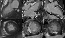
          A cohort of 40 consecutive children (age, 13.7 ± 3.3 years; 29 boys and 11 girls) with isolated LVNC underwent a baseline CMR scan with subsequent clinical follow-up. Short-axis cine images were used to calculate left ventricular (LV) ejection fraction (EF), end-diastolic volume (EDV), end-systolic volume (ESV), myocardial mass, ratio of non-compacted-to-compacted myocardial thickness (NC/C ratio), and number of non-compacted segments. The LGE images were analyzed to assess visually presence and patterns of LGE. The primary end point was a composite of cardiac death and heart transplantation.

          Results

          The LGE was present in 10 (25 %) children, and 46 (27 %) segments were involved, including 23 non-compacted segments and 23 normal segments. Compared with LGE- cohort, LGE+ cohort had significantly lower LVEF (23.8 ± 10.7 % vs. 42.9 ± 16.7 %, p < 0.001) and greater LVEDV (169.2 ± 65.1 vs. 118.2 ± 48.9 mL/m 2, p = 0.010), LVESV (131.3 ± 55.5 vs. 73.3 ± 46.7 mL/m 2, p = 0.002), and sphericity indices (0.75 ± 0.19 vs. 0.60 ± 0.20, p = 0.045). There were no differences in terms of number and distribution of non-compacted segments, NC/C ratio, and myocardial mass index between LGE+ and LGE- cohort. In the LGE+ cohort, adverse events occurred in 6 patients compared to 2 events in the LGE- cohort. Kaplan-Meier analysis showed a significant difference in outcome between LGE+ and LGE- cohort for cardiac death and heart transplantation ( p = 0.011).

          Conclusions

          The LGE was present in up to one-fourth of children with LVNC, and the LGE+ children exhibited a more maladaptive LV remodeling and a higher incidence of cardiovascular death and heart transplantation.

          Related collections

          Most cited references23

          • Record: found
          • Abstract: found
          • Article: not found

          Association of fibrosis with mortality and sudden cardiac death in patients with nonischemic dilated cardiomyopathy.

          Risk stratification of patients with nonischemic dilated cardiomyopathy is primarily based on left ventricular ejection fraction (LVEF). Superior prognostic factors may improve patient selection for implantable cardioverter-defibrillators (ICDs) and other management decisions. To determine whether myocardial fibrosis (detected by late gadolinium enhancement cardiovascular magnetic resonance [LGE-CMR] imaging) is an independent and incremental predictor of mortality and sudden cardiac death (SCD) in dilated cardiomyopathy. Prospective, longitudinal study of 472 patients with dilated cardiomyopathy referred to a UK center for CMR imaging between November 2000 and December 2008 after presence and extent of midwall replacement fibrosis were determined. Patients were followed up through December 2011. Primary end point was all-cause mortality. Secondary end points included cardiovascular mortality or cardiac transplantation; an arrhythmic composite of SCD or aborted SCD (appropriate ICD shock, nonfatal ventricular fibrillation, or sustained ventricular tachycardia); and a composite of HF death, HF hospitalization, or cardiac transplantation. Among the 142 patients with midwall fibrosis, there were 38 deaths (26.8%) vs 35 deaths (10.6%) among the 330 patients without fibrosis (hazard ratio [HR], 2.96 [95% CI, 1.87-4.69]; absolute risk difference, 16.2% [95% CI, 8.2%-24.2%]; P < .001) during a median follow-up of 5.3 years (2557 patient-years of follow-up). The arrhythmic composite was reached by 42 patients with fibrosis (29.6%) and 23 patients without fibrosis (7.0%) (HR, 5.24 [95% CI, 3.15-8.72]; absolute risk difference, 22.6% [95% CI, 14.6%-30.6%]; P < .001). After adjustment for LVEF and other conventional prognostic factors, both the presence of fibrosis (HR, 2.43 [95% CI, 1.50-3.92]; P < .001) and the extent (HR, 1.11 [95% CI, 1.06-1.16]; P < .001) were independently and incrementally associated with all-cause mortality. Fibrosis was also independently associated with cardiovascular mortality or cardiac transplantation (by fibrosis presence: HR, 3.22 [95% CI, 1.95-5.31], P < .001; and by fibrosis extent: HR, 1.15 [95% CI, 1.10-1.20], P < .001), SCD or aborted SCD (by fibrosis presence: HR, 4.61 [95% CI, 2.75-7.74], P < .001; and by fibrosis extent: HR, 1.10 [95% CI, 1.05-1.16], P < .001), and the HF composite (by fibrosis presence: HR, 1.62 [95% CI, 1.00-2.61], P = .049; and by fibrosis extent: HR, 1.08 [95% CI, 1.04-1.13], P < .001). Addition of fibrosis to LVEF significantly improved risk reclassification for all-cause mortality and the SCD composite (net reclassification improvement: 0.26 [95% CI, 0.11-0.41]; P = .001 and 0.29 [95% CI, 0.11-0.48]; P = .002, respectively). Assessment of midwall fibrosis with LGE-CMR imaging provided independent prognostic information beyond LVEF in patients with nonischemic dilated cardiomyopathy. The role of LGE-CMR in the risk stratification of dilated cardiomyopathy requires further investigation.
            Bookmark
            • Record: found
            • Abstract: found
            • Article: not found

            Isolated noncompaction of left ventricular myocardium. A study of eight cases.

            Isolated noncompaction of left ventricular myocardium is a rare disorder of endomyocardial morphogenesis characterized by numerous, excessively prominent ventricular trabeculations and deep intertrabecular recesses. This study comprised eight cases, including three at necropsy. Ages ranged from 11 months to 22.5 years, with follow-up as long as 5 years. Gross morphological severity ranged from moderately abnormal ventricular trabeculations to profoundly abnormal, loosely compacted trabeculations. Echocardiographic images were diagnostic and corresponded to the morphological appearances at necropsy. The depths of the intertrabecular recesses were assessed by a quantitative echocardiographic X-to-Y ratio and were significantly greater than in normal control subjects (p less than 0.001). Clinical manifestations of the disorder included depressed left ventricular systolic function in five patients, ventricular arrhythmias in five, systemic embolization in three, distinctive facial dysmorphism in three, and familial recurrence in four patients. We conclude that isolated noncompaction of left ventricular myocardium is a rare if not unique disorder with characteristic morphological features that can be identified by two-dimensional echocardiography. The incidence of cardiovascular complications is high. The disorder may be associated with facial dysmorphism and familial recurrence.
              Bookmark
              • Record: found
              • Abstract: found
              • Article: not found

              Long-term follow-up of 34 adults with isolated left ventricular noncompaction: a distinct cardiomyopathy with poor prognosis.

              We sought to describe characteristics and outcome in adults with isolated ventricular noncompaction (IVNC). Isolated ventricular noncompaction is an unclassified cardiomyopathy due to intrauterine arrest of compaction of the loose interwoven meshwork. Knowledge regarding diagnosis, morbidity and prognosis is limited. Echocardiographic criteria for IVNC include-in the absence of significant heart lesions-segmental thickening of the left ventricular myocardial wall consisting of two layers: a thin, compacted epicardial and an extremely thickened endocardial layer with prominent trabeculations and deep recesses. Thirty-four adults (age >16 years, 25 men) fulfilled the diagnostic criteria and were followed prospectively. At diagnosis, mean age was 42 + 17 years, and 12 patients (35%) were in New York Heart Association class III/IV. Left ventricular end-diastolic diameter was 65 + 12 mm and ejection fraction 33 + 13%. Apex and/or midventricular segments of both the inferior and lateral wall were involved in >80% of patients. Follow-up was 44 + 40 months. Major complications were heart failure in 18 patients (53%), thromboembolic events in 8 patients (24%) and ventricular tachycardias in 14 patients (41%). There were 12 deaths: sudden in six, end-stage heart failure in four and other causes in two patients. Four patients underwent heart transplantation. Automated cardioverter/defibrillators were implanted in four patients. Diagnosis of IVNC by echocardiography using strict criteria is feasible. Its mortality and morbidity are high, including heart failure, thrombo-embolic events and ventricular arrhythmias. Risk stratification includes heart failure therapy, oral anticoagulation, heart transplantation and implantation of an automated defibrillator/cardioverter. As IVNC is a distinct entity, its classification as a specific cardiomyopathy seems to be more appropriate.
                Bookmark

                Author and article information

                Contributors
                chenghb1981@163.com
                coolkan@163.com
                houch1968@sohu.com
                cheyne_xh@sina.com
                pathologyli@163.com
                doctorjing@126.com
                cardiacbmeyg@163.com
                cxy0202@126.com
                xlw6369@163.com
                Andrew41316@126.com
                fwyy06@126.com
                zsfuwai@vip.163.com
                S.Prasad@rbht.nhs.uk
                fuwaipujielin@163.com
                cjrzhaoshihua@163.com
                Journal
                J Cardiovasc Magn Reson
                J Cardiovasc Magn Reson
                Journal of Cardiovascular Magnetic Resonance
                BioMed Central (London )
                1097-6647
                1532-429X
                30 May 2015
                30 May 2015
                2015
                : 17
                : 1
                : 44
                Affiliations
                [ ]Center for Arrhythmia Diagnosis and Treatment, State Key Laboratory of Cardiovascular Disease, Fuwai Hospital, National Center for Cardiovascular Diseases, Chinese Academy of Medical Sciences and Peking Union Medical College, 100037 Beijing, People’s Republic of China
                [ ]Department of Radiology, State Key Laboratory of Cardiovascular Disease, Fuwai Hospital, National Center for Cardiovascular Diseases, Chinese Academy of Medical Sciences and Peking Union Medical College, 100037 Beijing, People’s Republic of China
                [ ]Department of Pathology, State Key Laboratory of Cardiovascular Disease, Fuwai Hospital, National Center for Cardiovascular Diseases, Chinese Academy of Medical Sciences and Peking Union Medical College, 100037 Beijing, People’s Republic of China
                [ ]Cardiovascular Magnetic Resonance Unit, NIHR Biomedical Research Unit, Royal Brompton Hospital, Sydney Street, SW3 6NP London, UK
                Article
                148
                10.1186/s12968-015-0148-7
                4449588
                26024839
                5f92ec4f-b3f5-41fe-8a10-7db672b8ee33
                © Cheng et al.; licensee BioMed Central. 2015

                This is an Open Access article distributed under the terms of the Creative Commons Attribution License ( http://creativecommons.org/licenses/by/4.0), which permits unrestricted use, distribution, and reproduction in any medium, provided the original work is properly credited. The Creative Commons Public Domain Dedication waiver ( http://creativecommons.org/publicdomain/zero/1.0/) applies to the data made available in this article, unless otherwise stated.

                History
                : 25 November 2014
                : 14 May 2015
                Categories
                Research
                Custom metadata
                © The Author(s) 2015

                Cardiovascular Medicine
                left ventricular non-compaction,cardiovascular magnetic resonance,late gadolinium enhancement,children

                Comments

                Comment on this article