8
views
0
recommends
+1 Recommend
0 collections
    0
    shares
      • Record: found
      • Abstract: found
      • Article: found
      Is Open Access

      The Role of the Pathogen Dose and PI3Kγ in Immunometabolic Reprogramming of Microglia for Innate Immune Memory

      research-article

      Read this article at

      Bookmark
          There is no author summary for this article yet. Authors can add summaries to their articles on ScienceOpen to make them more accessible to a non-specialist audience.

          Abstract

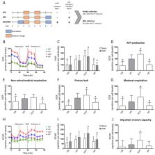
          Microglia, the innate immune cells of the CNS, exhibit long-term response changes indicative of innate immune memory (IIM). Our previous studies revealed IIM patterns of microglia with opposing immune phenotypes: trained immunity after a low dose and immune tolerance after a high dose challenge with pathogen-associated molecular patterns (PAMP). Compelling evidence shows that innate immune cells adopt features of IIM via immunometabolic control. However, immunometabolic reprogramming involved in the regulation of IIM in microglia has not been fully addressed. Here, we evaluated the impact of dose-dependent microglial priming with ultra-low (ULP, 1 fg/mL) and high (HP, 100 ng/mL) lipopolysaccharide (LPS) doses on immunometabolic rewiring. Furthermore, we addressed the role of PI3Kγ on immunometabolic control using naïve primary microglia derived from newborn wild-type mice, PI3Kγ-deficient mice and mice carrying a targeted mutation causing loss of lipid kinase activity. We found that ULP-induced IIM triggered an enhancement of oxygen consumption and ATP production. In contrast, HP was followed by suppressed oxygen consumption and glycolytic activity indicative of immune tolerance. PI3Kγ inhibited glycolysis due to modulation of cAMP-dependent pathways. However, no impact of specific PI3Kγ signaling on immunometabolic rewiring due to dose-dependent LPS priming was detected. In conclusion, immunometabolic reprogramming of microglia is involved in IIM in a dose-dependent manner via the glycolytic pathway, oxygen consumption and ATP production: ULP (ultra-low-dose priming) increases it, while HP reduces it.

          Related collections

          Most cited references64

          • Record: found
          • Abstract: found
          • Article: not found

          Analyzing real-time PCR data by the comparative C(T) method.

          Two different methods of presenting quantitative gene expression exist: absolute and relative quantification. Absolute quantification calculates the copy number of the gene usually by relating the PCR signal to a standard curve. Relative gene expression presents the data of the gene of interest relative to some calibrator or internal control gene. A widely used method to present relative gene expression is the comparative C(T) method also referred to as the 2 (-DeltaDeltaC(T)) method. This protocol provides an overview of the comparative C(T) method for quantitative gene expression studies. Also presented here are various examples to present quantitative gene expression data using this method.
            Bookmark
            • Record: found
            • Abstract: found
            • Article: not found

            Resting microglial cells are highly dynamic surveillants of brain parenchyma in vivo.

            Microglial cells represent the immune system of the mammalian brain and therefore are critically involved in various injuries and diseases. Little is known about their role in the healthy brain and their immediate reaction to brain damage. By using in vivo two-photon imaging in neocortex, we found that microglial cells are highly active in their presumed resting state, continually surveying their microenvironment with extremely motile processes and protrusions. Furthermore, blood-brain barrier disruption provoked immediate and focal activation of microglia, switching their behavior from patroling to shielding of the injured site. Microglia thus are busy and vigilant housekeepers in the adult brain.
              Bookmark
              • Record: found
              • Abstract: found
              • Article: not found

              Succinate is an inflammatory signal that induces IL-1β through HIF-1α.

              Macrophages activated by the Gram-negative bacterial product lipopolysaccharide switch their core metabolism from oxidative phosphorylation to glycolysis. Here we show that inhibition of glycolysis with 2-deoxyglucose suppresses lipopolysaccharide-induced interleukin-1β but not tumour-necrosis factor-α in mouse macrophages. A comprehensive metabolic map of lipopolysaccharide-activated macrophages shows upregulation of glycolytic and downregulation of mitochondrial genes, which correlates directly with the expression profiles of altered metabolites. Lipopolysaccharide strongly increases the levels of the tricarboxylic-acid cycle intermediate succinate. Glutamine-dependent anerplerosis is the principal source of succinate, although the 'GABA (γ-aminobutyric acid) shunt' pathway also has a role. Lipopolysaccharide-induced succinate stabilizes hypoxia-inducible factor-1α, an effect that is inhibited by 2-deoxyglucose, with interleukin-1β as an important target. Lipopolysaccharide also increases succinylation of several proteins. We therefore identify succinate as a metabolite in innate immune signalling, which enhances interleukin-1β production during inflammation.
                Bookmark

                Author and article information

                Contributors
                Role: Academic Editor
                Journal
                Int J Mol Sci
                Int J Mol Sci
                ijms
                International Journal of Molecular Sciences
                MDPI
                1422-0067
                04 March 2021
                March 2021
                : 22
                : 5
                : 2578
                Affiliations
                [1 ]Institute of Molecular Cell Biology, Jena University Hospital, D-07745 Jena, Germany; trim.lajqi@ 123456med.uni-heidelberg.de (T.L.); fabienne.haas@ 123456med.uni-jena.de (F.H.); grosse_silke@ 123456gmx.de (S.G.); regine.heller@ 123456med.uni-jena.de (R.H.)
                [2 ]Department of Neonatology, Heidelberg University Children’s Hospital, D-69120 Heidelberg, Germany; hannes.hudalla@ 123456med.uni-heidelberg.de
                [3 ]Leibniz Institute on Ageing, Fritz Lipmann Institute, D-07745 Jena, Germany; christian.marx@ 123456leibniz-fli.de (C.M.); zhao-qi.wang@ 123456leibniz-fli.de (Z.-Q.W.)
                [4 ]Department of Anesthesiology and Intensive Care Medicine, Jena University Hospital, D-07747 Jena, Germany; michael.bauer@ 123456med.uni-jena.de (M.B.); reinhard.wetzker@ 123456uni-jena.de (R.W.)
                Author notes
                [* ]Correspondence: reinhard.bauer@ 123456med.uni-jena.de ; Tel.: +49-364-1939-5636; Fax: +49-364-1939-5602
                Author information
                https://orcid.org/0000-0002-2584-589X
                https://orcid.org/0000-0002-8336-3485
                Article
                ijms-22-02578
                10.3390/ijms22052578
                7961448
                33806610
                612e236c-c09a-4538-8e0b-ca53686588db
                © 2021 by the authors.

                Licensee MDPI, Basel, Switzerland. This article is an open access article distributed under the terms and conditions of the Creative Commons Attribution (CC BY) license ( http://creativecommons.org/licenses/by/4.0/).

                History
                : 29 December 2020
                : 26 February 2021
                Categories
                Article

                Molecular biology
                microglia,immunometabolism,innate immune memory,lps,pi3kγ,glycolysis,pentose phosphate pathway,oxphos,ocr,ecar

                Comments

                Comment on this article