1
views
0
recommends
+1 Recommend
0 collections
    0
    shares
      • Record: found
      • Abstract: found
      • Article: found
      Is Open Access

      Tanshinone IIA protects mice against atherosclerotic injury by activating the TGF-β/PI3K/Akt/eNOS pathway

      research-article

      Read this article at

      Bookmark
          There is no author summary for this article yet. Authors can add summaries to their articles on ScienceOpen to make them more accessible to a non-specialist audience.

          Objective

          Explored the mechanism of action of tanshinone IIA (TIIA) against atherosclerosis.

          Methods

          ApoE −/− mice were divided into two groups of 10: model and TIIA. A control group of 10 wild-type mice was created. ApoE −/− mice were fed a high-fat diet for 12 weeks. The TIIA group received TIIA once daily. Mice were anesthetized, blood collected by cardiac puncture, and the aortic sinus/arch collected for histology and molecular studies, respectively.

          Results

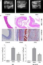
          Mice intima in the model group had large areas of plaque formation, serum levels of total cholesterol (TC), triglycerides, and low-density lipoprotein-cholesterol (LDL-C) increased significantly, and high-density lipoprotein-cholesterol (HDL-C) levels decreased significantly in the model group after 12 weeks. Staining [hematoxylin and eosin (H&E), Oil-Red-O] showed that the aorta had lesions, a higher degree of plaque formation, and considerable lipid deposition in model-group mice. After TIIA treatment, expression of HDL-C was increased significantly and that of TC, triglycerides and LDL-C decreased significantly, and plaque size and lipid deposition improved obviously. Analyses of protein phosphorylation in aortic tissue suggested that the transforming growth factor (TGF)-β/phosphatidylinositol 3-kinase (PI3K)/protein kinase B (Akt)/endothelial nitric oxide synthase (eNOS) pathway was activated in TIIA-treated mice.

          Conclusion

          TIIA can lower levels of serum lipids, stabilize atherosclerotic plaques, reduce endothelial injury, and inflammatory damage by activation of the TGF-β/PI3K/Akt/eNOS pathway.

          Related collections

          Most cited references27

          • Record: found
          • Abstract: found
          • Article: found
          Is Open Access

          The Role of Mammalian Target of Rapamycin (mTOR) in Insulin Signaling

          The mammalian target of rapamycin (mTOR) is a serine/threonine kinase that controls a wide spectrum of cellular processes, including cell growth, differentiation, and metabolism. mTOR forms two distinct multiprotein complexes known as mTOR complex 1 (mTORC1) and mTOR complex 2 (mTORC2), which are characterized by the presence of raptor and rictor, respectively. mTOR controls insulin signaling by regulating several downstream components such as growth factor receptor-bound protein 10 (Grb10), insulin receptor substrate (IRS-1), F-box/WD repeat-containing protein 8 (Fbw8), and insulin like growth factor 1 receptor/insulin receptor (IGF-IR/IR). In addition, mTORC1 and mTORC2 regulate each other through a feedback loop to control cell growth. This review outlines the current understanding of mTOR regulation in insulin signaling in the context of whole body metabolism.
            Bookmark
            • Record: found
            • Abstract: found
            • Article: not found

            Hypoxia inducible factor as a therapeutic target for atherosclerosis.

            Atherosclerosis is a highly prevalent disease that can significantly increase the risk of major vascular events, such as myocardial or cerebral infarctions. The anoxemia theory states that a disparity between oxygen supply and demand contributes to atherosclerosis. Hypoxia inducible factor-1 (HIF-1) is a heterodimeric protein, part of the basic helix-loop-helix family and one of the main regulators of cellular responses in a low‑oxygen environment. It plays a key role in the development of atherosclerosis through cell-specific responses, acting on endothelial cells, vascular smooth muscle cells (SMCs) and macrophages. Through the upregulation of VEGF, NO, ROS and PDGF, HIF-1 is able to cause endothelial cell dysfunction, proliferation, angiogenesis and inflammation. Activation of the NF-kB pathway in endothelial cells is an important contributor to inflammation and positively feedbacks to HIF-1. HIF-1 also plays a significant role in both the proliferation and migration of smooth muscle cells - two important features of atherosclerosis, while the formation of foam cells (lipid-laden macrophages) is also a critical step in atherosclerosis and mediated by HIF-1 through various mechanisms such as dysfunctional efflux pathways in macrophages. Overall, HIF-1 exerts its effect on the pathogenesis of atherosclerosis via a variety of molecular and cellular events in the process. In this review article, we examine the effects HIF-1 on vascular cells and macrophages in the development of atherosclerosis, highlighting the environmental cues and signalling pathways that control HIF-1 expression/activation within the vasculature. We will highlight the potential of HIF-1 as a therapeutic target on the disease development.
              Bookmark
              • Record: found
              • Abstract: found
              • Article: not found

              CD40 ligand mediates inflammation independently of CD40 by interaction with Mac-1.

              Strong evidence supports a role for CD40 ligand (CD40L) as marker and mediator of inflammatory diseases such as atherosclerosis. Despite extensive characterization of CD40, the classic receptor of CD40L, its role in immune defense against inflammatory diseases remains uncertain. The present study aimed to characterize the contribution of CD40 signaling to atherogenesis. Surprisingly, mice deficient in both CD40 and the low-density lipoprotein receptor did not develop smaller lesions in the aortic arch, root, and thoracoabdominal aorta compared with mice deficient only in the low-density lipoprotein receptor that consumed an atherogenic diet for 8 and 16 weeks. By flow cytometry, radioactive binding assays, and immunoprecipitation, we demonstrate that CD40L interacts with the integrin Mac-1, which results in Mac-1-dependent adhesion and migration of inflammatory cells as well as myeloperoxidase release in vitro. Furthermore, mice deficient in CD40L show significantly reduced thioglycolate-elicited invasion of inflammatory cells into the peritoneal cavity compared with mice deficient in CD40 and wild-type controls. Inhibition of Mac-1 in low-density lipoprotein receptor-deficient mice attenuates lesion development and reduces lesional macrophage accumulation. These observations identify the interaction of CD40L and Mac-1 as an alternative pathway for CD40L-mediated inflammation. This novel mechanism expands understanding of inflammatory signaling during atherogenesis.
                Bookmark

                Author and article information

                Journal
                Coron Artery Dis
                Coron. Artery Dis
                CAD
                Coronary Artery Disease
                Lippincott Williams & Wilkins
                0954-6928
                1473-5830
                June 2020
                13 December 2019
                : 31
                : 4
                : 385-392
                Affiliations
                [a ]Key Laboratory of Ministry of Education for TCM Viscera-State Theory and Applications
                [b ]Department of Medical Science of Laboratory
                [c ]Basic Medical College, Liaoning University of Traditional Chinese Medicine, Shenyang, Liaoning, China
                Author notes
                Correspondence to Lianqun Jia, PhD, Key Laboratory of Ministry of Education for TCM Viscera-State Theory and Applications, Liaoning University of Traditional Chinese Medicine, No. 79, Chongshan Eastern Road, Shenyang, Liaoning 110847, China, Tel: +86 0243 1207 185; fax: +86 0243 1207 358; e-mail: jlq-8@ 123456163.com yang_guanlin@ 123456163.com
                Article
                00010
                10.1097/MCA.0000000000000835
                7192539
                31842027
                63102c56-4bda-4b10-86c3-434feb7c4677
                Copyright © 2019 The Author(s). Published by Wolters Kluwer Health, Inc.

                This is an open-access article distributed under the terms of the Creative Commons Attribution-Non Commercial-No Derivatives License 4.0 (CCBY-NC-ND), where it is permissible to download and share the work provided it is properly cited. The work cannot be changed in any way or used commercially without permission from the journal.

                History
                : 3 January 2019
                : 10 November 2019
                Categories
                Translational Science
                Custom metadata
                TRUE

                atherosclerosis,endothelial nitric oxide synthase,phosphatidylinositol 3-kinase/protein kinase b,tanshinone iia,transforming growth factor β

                Comments

                Comment on this article