9
views
0
recommends
+1 Recommend
0 collections
    0
    shares
      • Record: found
      • Abstract: found
      • Article: found
      Is Open Access

      Identification of Key Residues for Urate Specific Transport in Human Glucose Transporter 9 (hSLC2A9)

      research-article

      Read this article at

      Bookmark
          There is no author summary for this article yet. Authors can add summaries to their articles on ScienceOpen to make them more accessible to a non-specialist audience.

          Abstract

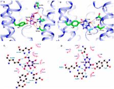
          Human glucose transporter 9 (hSLC2A9) is critical in human urate homeostasis, for which very small deviations can lead to chronic or acute metabolic disorders. Human SLC2A9 is unique in that it transports hexoses as well as the organic anion, urate. This ability is in contrast to other homologous sugar transporters such as glucose transporters 1 and 5 (SLC2A1 & SLC2A5) and the xylose transporter (XylE), despite the fact that these transporters have similar protein structures. Our in silico substrate docking study has revealed that urate and fructose bind within the same binding pocket in hSLC2A9, yet with distinct orientations, and allowed us to identify novel residues for urate binding. Our functional studies confirmed that N429 is a key residue for both urate binding and transport. We have shown that cysteine residues, C181, C301 and C459 in hSLC2A9 are also essential elements for mediating urate transport. Additional data from chimæric protein analysis illustrated that transmembrane helix 7 of hSLC2A9 is necessary for urate transport but not sufficient to allow urate transport to be induced in glucose transporter 5 (hSLC2A5). These data indicate that urate transport in hSLC2A9 involves several structural elements rather than just a unique substrate binding pocket.

          Related collections

          Most cited references31

          • Record: found
          • Abstract: found
          • Article: not found

          Crystal structure of the human glucose transporter GLUT1.

          The glucose transporter GLUT1 catalyses facilitative diffusion of glucose into erythrocytes and is responsible for glucose supply to the brain and other organs. Dysfunctional mutations may lead to GLUT1 deficiency syndrome, whereas overexpression of GLUT1 is a prognostic indicator for cancer. Despite decades of investigation, the structure of GLUT1 remains unknown. Here we report the crystal structure of human GLUT1 at 3.2 Å resolution. The full-length protein, which has a canonical major facilitator superfamily fold, is captured in an inward-open conformation. This structure allows accurate mapping and potential mechanistic interpretation of disease-associated mutations in GLUT1. Structure-based analysis of these mutations provides an insight into the alternating access mechanism of GLUT1 and other members of the sugar porter subfamily. Structural comparison of the uniporter GLUT1 with its bacterial homologue XylE, a proton-coupled xylose symporter, allows examination of the transport mechanisms of both passive facilitators and active transporters.
            Bookmark
            • Record: found
            • Abstract: found
            • Article: not found

            SLC2A9 is a newly identified urate transporter influencing serum urate concentration, urate excretion and gout.

            Uric acid is the end product of purine metabolism in humans and great apes, which have lost hepatic uricase activity, leading to uniquely high serum uric acid concentrations (200-500 microM) compared with other mammals (3-120 microM). About 70% of daily urate disposal occurs via the kidneys, and in 5-25% of the human population, impaired renal excretion leads to hyperuricemia. About 10% of people with hyperuricemia develop gout, an inflammatory arthritis that results from deposition of monosodium urate crystals in the joint. We have identified genetic variants within a transporter gene, SLC2A9, that explain 1.7-5.3% of the variance in serum uric acid concentrations, following a genome-wide association scan in a Croatian population sample. SLC2A9 variants were also associated with low fractional excretion of uric acid and/or gout in UK, Croatian and German population samples. SLC2A9 is a known fructose transporter, and we now show that it has strong uric acid transport activity in Xenopus laevis oocytes.
              Bookmark
              • Record: found
              • Abstract: found
              • Article: not found

              Crystal structure of a bacterial homologue of glucose transporters GLUT1-4.

              Glucose transporters are essential for metabolism of glucose in cells of diverse organisms from microbes to humans, exemplified by the disease-related human proteins GLUT1, 2, 3 and 4. Despite rigorous efforts, the structural information for GLUT1-4 or their homologues remains largely unknown. Here we report three related crystal structures of XylE, an Escherichia coli homologue of GLUT1-4, in complex with d-xylose, d-glucose and 6-bromo-6-deoxy-D-glucose, at resolutions of 2.8, 2.9 and 2.6 Å, respectively. The structure consists of a typical major facilitator superfamily fold of 12 transmembrane segments and a unique intracellular four-helix domain. XylE was captured in an outward-facing, partly occluded conformation. Most of the important amino acids responsible for recognition of D-xylose or d-glucose are invariant in GLUT1-4, suggesting functional and mechanistic conservations. Structure-based modelling of GLUT1-4 allows mapping and interpretation of disease-related mutations. The structural and biochemical information reported here constitutes an important framework for mechanistic understanding of glucose transporters and sugar porters in general.
                Bookmark

                Author and article information

                Journal
                Sci Rep
                Sci Rep
                Scientific Reports
                Nature Publishing Group
                2045-2322
                24 January 2017
                2017
                : 7
                : 41167
                Affiliations
                [1 ]Department of Physiology, Faculty of Medicine and Dentistry, University of Alberta, Alberta , Canada
                [2 ]Department of Biochemistry, Faculty of Medicine and Dentistry, University of Alberta, Alberta , Canada
                Author notes
                Article
                srep41167
                10.1038/srep41167
                5259734
                28117388
                64e06390-df8f-4532-809f-e287d5bbedde
                Copyright © 2017, The Author(s)

                This work is licensed under a Creative Commons Attribution 4.0 International License. The images or other third party material in this article are included in the article’s Creative Commons license, unless indicated otherwise in the credit line; if the material is not included under the Creative Commons license, users will need to obtain permission from the license holder to reproduce the material. To view a copy of this license, visit http://creativecommons.org/licenses/by/4.0/

                History
                : 22 September 2016
                : 14 December 2016
                Categories
                Article

                Uncategorized
                Uncategorized

                Comments

                Comment on this article