5
views
0
recommends
+1 Recommend
0 collections
    0
    shares
      • Record: found
      • Abstract: found
      • Article: found
      Is Open Access

      Utility of virtual monoenergetic images from spectral detector computed tomography in improving image segmentation for purposes of 3D printing and modeling

      research-article

      Read this article at

      Bookmark
          There is no author summary for this article yet. Authors can add summaries to their articles on ScienceOpen to make them more accessible to a non-specialist audience.

          Abstract

          Background

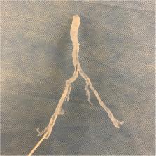
          One of the key steps in generating three-dimensional (3D) printed models in medicine is segmentation of radiologic imaging. The software tools used for segmentation may be automated, semi-automated, or manual which rely on differences in material density, attenuation characteristics, and/or advanced software algorithms. Spectral Detector Computed Tomography (SDCT) is a form of dual energy computed tomography that works at the detector level to generate virtual monoenergetic images (VMI) at different energies/ kilo-electron volts (keV). These VMI have varying contrast and attenuation characteristics relative to material density. The purpose of this pilot project is to explore the use of VMI in segmentation for medical 3D printing in four separate clinical scenarios. Cases were retrospectively selected based on varying complexity, value of spectral data, and across multiple clinical disciplines (Vascular, Cardiology, Oncology, and Orthopedic).

          Results

          In all four clinical cases presented, the segmentation process was qualitatively reported as easier, faster, and increased the operator’s confidence in obtaining accurate anatomy. All cases demonstrated a significant difference in the calculated Hounsfield Units between conventional and VMI data at the level of targeted segmentation anatomy. Two cases would not have been feasible for segmentation and 3D printing using conventional images only. VMI data significantly reduced conventional CT artifacts in one of the cases.

          Conclusion

          Utilization of VMI from SDCT can improve and assist the segmentation of target anatomy for medical 3D printing by enhancing material contrast and decreasing CT artifact.

          Electronic supplementary material

          The online version of this article (10.1186/s41205-019-0038-y) contains supplementary material, which is available to authorized users.

          Related collections

          Most cited references15

          • Record: found
          • Abstract: found
          • Article: not found

          Dual- and Multi-Energy CT: Principles, Technical Approaches, and Clinical Applications.

          In x-ray computed tomography (CT), materials having different elemental compositions can be represented by identical pixel values on a CT image (ie, CT numbers), depending on the mass density of the material. Thus, the differentiation and classification of different tissue types and contrast agents can be extremely challenging. In dual-energy CT, an additional attenuation measurement is obtained with a second x-ray spectrum (ie, a second "energy"), allowing the differentiation of multiple materials. Alternatively, this allows quantification of the mass density of two or three materials in a mixture with known elemental composition. Recent advances in the use of energy-resolving, photon-counting detectors for CT imaging suggest the ability to acquire data in multiple energy bins, which is expected to further improve the signal-to-noise ratio for material-specific imaging. In this review, the underlying motivation and physical principles of dual- or multi-energy CT are reviewed and each of the current technical approaches is described. In addition, current and evolving clinical applications are introduced.
            Bookmark
            • Record: found
            • Abstract: found
            • Article: not found

            Medical 3D Printing for the Radiologist.

            While use of advanced visualization in radiology is instrumental in diagnosis and communication with referring clinicians, there is an unmet need to render Digital Imaging and Communications in Medicine (DICOM) images as three-dimensional (3D) printed models capable of providing both tactile feedback and tangible depth information about anatomic and pathologic states. Three-dimensional printed models, already entrenched in the nonmedical sciences, are rapidly being embraced in medicine as well as in the lay community. Incorporating 3D printing from images generated and interpreted by radiologists presents particular challenges, including training, materials and equipment, and guidelines. The overall costs of a 3D printing laboratory must be balanced by the clinical benefits. It is expected that the number of 3D-printed models generated from DICOM images for planning interventions and fabricating implants will grow exponentially. Radiologists should at a minimum be familiar with 3D printing as it relates to their field, including types of 3D printing technologies and materials used to create 3D-printed anatomic models, published applications of models to date, and clinical benefits in radiology. Online supplemental material is available for this article.
              Bookmark
              • Record: found
              • Abstract: found
              • Article: not found

              Dual-energy CT: general principles.

              In dual-energy CT (DECT), two CT datasets are acquired with different x-ray spectra. These spectra are generated using different tube potentials, partially also with additional filtration at 140 kVp. Spectral information can also be resolved by layer detectors or quantum-counting detectors. Several technical approaches-that is, sequential acquisition, rapid voltage switching, dual-source CT (DSCT), layer detector, quantum-counting detector-offer different spectral contrast and dose efficiency. Various postprocessing algorithms readily provide clinically relevant spectral information.
                Bookmark

                Author and article information

                Contributors
                egk8@case.edu
                nils.grosse-hokamp@uk-koeln.de
                Leslie.Ciancibello@UHhospitals.org
                Nikhil.Ramaiya@uhhospitals.org
                Christos.Kosmas@UHhospitals.org
                Amit.Gupta@UHhospitals.org
                Journal
                3D Print Med
                3D Print Med
                3D Printing in Medicine
                Springer International Publishing (Cham )
                2365-6271
                18 January 2019
                18 January 2019
                December 2019
                : 5
                : 1
                Affiliations
                [1 ]ISNI 0000 0000 9149 4843, GRID grid.443867.a, Department of Radiology, , University Hospitals Cleveland Medical Center/Case Western Reserve University, ; 11100 Euclid Ave, Cleveland, OH 44106 USA
                [2 ]ISNI 0000 0000 8852 305X, GRID grid.411097.a, Institute for Diagnostic and Interventional Radiology, , University Hospital Cologne, ; Cologne, Germany
                Author information
                http://orcid.org/0000-0002-8932-3433
                Article
                38
                10.1186/s41205-019-0038-y
                6505638
                30659415
                6678707b-bebf-4796-a781-7fca42bebd0a
                © The Author(s) 2019

                Open AccessThis article is distributed under the terms of the Creative Commons Attribution 4.0 International License ( http://creativecommons.org/licenses/by/4.0/), which permits unrestricted use, distribution, and reproduction in any medium, provided you give appropriate credit to the original author(s) and the source, provide a link to the Creative Commons license, and indicate if changes were made.

                History
                : 20 September 2018
                : 3 January 2019
                Categories
                Research
                Custom metadata
                © The Author(s) 2019

                3d printing,spectral detector ct,segmentation,dual-energy ct,dual layer ct

                Comments

                Comment on this article