31
views
0
recommends
+1 Recommend
0 collections
    0
    shares
      • Record: found
      • Abstract: found
      • Article: not found

      Zoonotic and Non-zoonotic Parasites of Wild Rodents in Turkman Sahra, Northeastern Iran

      research-article

      Read this article at

      ScienceOpenPMC
      Bookmark
          There is no author summary for this article yet. Authors can add summaries to their articles on ScienceOpen to make them more accessible to a non-specialist audience.

          Abstract

          Background:

          This study was conducted to collect informative data on the parasitic infection of wild rodents, emphasizing on finding parasites, which have medical importance to human.

          Methods:

          During 2012–2014, a total number of 91 wild rodents were captured from rural areas of Turkmen Sahra, Golestan Province, using handmade traps. Animals were anesthetized, surveyed for any ectoparasite and then their carcasses were carefully dissected for examination of endoparsites.

          Results:

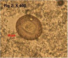
          Four species of rodents including Mus musculus (52.75%), Rattus norvegicus (38.46%), Rhombomys opimus (4.40%) and Meriones libycus (4.40%) were captured. Parasitic infestation was detected in 38.5% of sampled rodents. Parasite infestation rates of sampled rodents was Hymenolepis diminuta = 7.7%, Cryptosporidium spp = 6.6%, Trichuris spp.= 5.5%, Cysticercus fasciolaris = 2.20%, Angiostrongylus spp.= 2.20%, Capillaria sp.= 1.09%, Rhipicephalus spp. = 8.70%, Nosopsyllus fasciatus = 1.09%, and Laelaps nuttalli = 3.29%. Among 10 genera/species of identified parasites, at least 8 of them were zoonotic with public health importance. L. nuttalli and N. fasciatus were the only two non-zoonotic detected parasites in this survey.

          Conclusion:

          Harboring a wide variety of zoonotic parasites in sampled wild rodents particularly when they live nearby villages, represents a potential risk to native inhabitants. Hence, controlling rodents’ population in residential regions and improving awareness of local people about the risk of disease transmission through rodents seems to be entirely necessary.

          Related collections

          Most cited references9

          • Record: found
          • Abstract: found
          • Article: not found

          The present status of human helminthic diseases in Iran.

          M Rokni (2008)
          Over the last few decades there have been several marked changes in the human helminthiases found in Iran. Fascioliasis is emerging as an important chronic disease of humans, especially in the northern province of Gilan (where outbreaks in 1989 and 1999 involved >7000 and >10,000 cases, respectively) and, more recently, in the western province of Kermanshah. In contrast, no cases of urinary schistosomiasis, a disease that once affected thousands of individuals in south-western Khuzestan province, have been reported in Iran in recent years, and no cases of dracunculiasis have been seen in the country since the mid-1970s. Approximately 1% of all admissions to surgical wards are attributable to cystic echinococcosis, which is still considered endemic, but only a few cases of alveolar echinococcosis have been recorded. Over the last decade, there appears to have been a generally downward trend in the incidence of intestinal helminthiases in Iran. Recent estimates of the prevalences of ascariasis and strongyloidiasis, for example, lie between just 0.1% and 0.3%, and <1% of the population now appears to be infected with hookworm. In contrast, human infection with Hymenolepis and Enterobius remains relatively common. There have been a few case reports of toxocariasis and a few sero-epidemiological investigations of this disease but problems in accurate diagnosis have prevented good estimates of the general prevalence of this nematode infection. Just nine cases of pentastomiasis (all caused by Linguatula), 12 of dirofilariasis, one of gongylonemiasis, and three of moniliformiasis have been formally recorded in Iran.
            Bookmark
            • Record: found
            • Abstract: found
            • Article: found
            Is Open Access

            Endoparasites of Rodents and Their Zoonotic Importance in Germi, Dashte–Mogan, Ardabil Province, Iran

            Background In order to verify the infectivity of rodents with endoparasites in Germi (Dashte-Mogan, Ardabil Province) the current study was undertaken. Methods Using live traps, 177 rodents were trapped during 2005–2007. In field laboratory, all rodents were bled prior to autopsy, frozen at −20°C, and shipped to the School of Public Health, Tehran University of Medical Sciences, Iran. In parasitological laboratory, every rodent was dissected and its different organs were examined for the presence of any parasite. Blood thick and thin smears as well as impression smears of liver and spleen were stained with Geimsa and examined microscopically. Results Two species of rodents were trapped; Meriones persicus (90.4%) and Microtus socialis (9.6%). The species of parasites found in M. persicus and their prevalences were as follows: Hymenolepis diminuta (38.8%), Hymenolepis nana (2.5%), Trichuris sp.(40.6), Mesocestoides larva (=tetrathyridium) (3.1%), Capillaria hepatica (6.9%), Moniliformis moniliformis (11.3%), Syphacia obvelata (2.5%), Taenia endothoracicus larva (0.6%), Physaloptera sp. (0.6%), Dentostomella translucida (0.6%), Heligmosomum mixtum (0.6%), Strobilocercus fasciolaris (0.6%),and Aspiculuris tetraptera (0.6%). The species of parasites found in M. socialis and their prevalences were as follows: H. diminuta (17.6%), Trichuris sp. (5.9%), Mesocestoides larva (5.9%), S. obvelata (11.8%), S. syphacia (11.8%), H. mixtum (17.6%), and Aspiculuris tetraptera (11.8%). There were no statistical differences between male and female for infectivity with parasites in either M. persicus or M. socialis. No blood or tissue protozoan parasite was found in any of the rodents examined. Conclusion Among different species identified, some had zoonotic importance. Therefore, the potential health hazard of these species needs to be considered to prevent infectivity of humans.
              Bookmark
              • Record: found
              • Abstract: found
              • Article: not found

              Development of a multilocus sequence tool for typing Cryptosporidium muris and Cryptosporidium andersoni.

              Although widely used for the characterization of the transmission of intestinal Cryptosporidium spp., genotyping tools are not available for C. muris and C. andersoni, two of the most common gastric Cryptosporidium spp. infecting mammals. In this study, we screened the C. muris whole-genome sequencing data for microsatellite and minisatellite sequences. Among the 13 potential loci (6 microsatellite and 7 minisatellite loci) evaluated by PCR and DNA sequencing, 4 were eventually chosen. DNA sequence analyses of 27 C. muris and 17 C. andersoni DNA preparations showed the presence of 5 to 10 subtypes of C. muris and 1 to 4 subtypes of C. andersoni at each locus. Altogether, 11 C. muris and 7 C. andersoni multilocus sequence typing (MLST) subtypes were detected among the 16 C. muris and 12 C. andersoni specimens successfully sequenced at all four loci. In all analyses, the C. muris isolate (TS03) that originated from an East African mole rat differed significantly from other C. muris isolates, approaching the extent of genetic differences between C. muris and C. andersoni. Thus, an MLST technique was developed for the high-resolution typing of C. muris and C. andersoni. It should be useful for the characterization of the population genetics and transmission of gastric Cryptosporidium spp.
                Bookmark

                Author and article information

                Journal
                Iran J Parasitol
                Iran J Parasitol
                IJPA
                IJPA
                Iranian Journal of Parasitology
                Tehran University of Medical Sciences
                1735-7020
                2008-238X
                Jul-Sep 2016
                : 11
                : 3
                : 350-357
                Affiliations
                [1. ]Faculty of Fisheries and Environment, Gorgan University of Agricultural Sciences and Natural Resources, Gorgan, Iran
                [2. ]Department of Parasitology Faculty of Veterinary Medicine University of Tehran, Tehran, Iran
                Author notes
                [* ] Correspondence Email: snamroodi2000@ 123456yahoo.com
                Article
                ijpa-11-350
                5256051
                28127340
                6b4efaab-229d-429b-bacc-e89290abc910
                Copyright© Iranian Society of Parasitology & Tehran University of Medical Sciences

                This work is licensed under a Creative Commons Attribution-NonCommercial 3.0 Unported License which allows users to read, copy, distribute and make derivative works for non-commercial purposes from the material, as long as the author of the original work is cited properly.

                History
                : 21 February 2015
                : 12 June 2016
                Categories
                Original Article

                Parasitology
                rodents,zoonotic parasite,non-zoonotic parasite,iran
                Parasitology
                rodents, zoonotic parasite, non-zoonotic parasite, iran

                Comments

                Comment on this article