6
views
0
recommends
+1 Recommend
0 collections
    0
    shares
      • Record: found
      • Abstract: found
      • Article: found
      Is Open Access

      Diagnostic accuracy of endoscopy for the detection of isthmuses of mandibular molar teeth using micro-CT as reference

      research-article

      Read this article at

      Bookmark
          There is no author summary for this article yet. Authors can add summaries to their articles on ScienceOpen to make them more accessible to a non-specialist audience.

          Abstract

          Purpose:

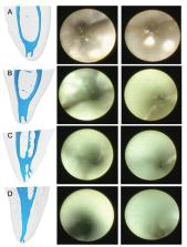
          The aim of this study was to evaluate the efficacy of endoscopic visualisation to detect the presence and type of isthmuses within the mesial root canals of mandibular first molar teeth compared with micro-computed tomography (micro- CT) images as reference.

          Materials and methods:

          Thirty-two mesial roots of mandibular first molars presenting isthmuses were selected based on micro-CT scans. In all, 12 type I and 20 band-shaped isthmuses were collected. The specimens were mounted in the posterior socket of dental phantom manikin for endoscopic visualisation. The ability of endoscopes to visualize the presence of isthmuses and distinguish the type of isthmuses was compared. Micro-CT images of the specimens were used as references. Data were analyzed using Fisher’s exact tests.

          Results:

          Sensitivity of endoscope to detect isthmuses were also calculated for each isthmus type. In 37.5% of the samples, isthmus presence was correctly diagnosed via orthograde endoscopic visualization. Type I istmuses were significantly more detected than band-shaped isthmuses (P<0.05). Endoscope showed higher sensitivity to detect type I isthmus than band-shaped isthmus.

          Conclusion:

          The endodontic endoscope could detect type I isthmuses more accurately than band- shaped isthmuses.

          Related collections

          Most cited references23

          • Record: found
          • Abstract: found
          • Article: not found

          Comparative accuracy of the Clearing Technique, CBCT and Micro-CT methods in studying the mesial root canal configuration of mandibular first molars.

          To compare the accuracy of the clearing technique and cone beam computed tomography (CBCT) in the assessment of root canal configurations using micro-computed tomography (micro-CT) imaging system as the reference standard.
            Bookmark
            • Record: found
            • Abstract: found
            • Article: not found

            Correlative bacteriologic and micro-computed tomographic analysis of mandibular molar mesial canals prepared by self-adjusting file, reciproc, and twisted file systems.

            This ex vivo study evaluated the disinfecting and shaping ability of 3 protocols used in the preparation of mesial root canals of mandibular molars by means of correlative bacteriologic and micro-computed tomographic (μμCT) analysis.
              Bookmark
              • Record: found
              • Abstract: found
              • Article: not found

              Frequency and type of canal isthmuses in first molars detected by endoscopic inspection during periradicular surgery.

              T von Arx (2005)
              To analyse the occurrence of canal isthmuses in molars following root-end resection.
                Bookmark

                Author and article information

                Journal
                Eur Oral Res
                Eur Oral Res
                eor
                eor
                IUFD
                European Oral Research
                Istanbul University Faculty of Dentistry (Istanbul, Turkey )
                2630-6158
                2651-2823
                04 January 2021
                04 January 2021
                : 55
                : 1
                : 34-38
                Affiliations
                [1 ]Department of Endodontics, Faculty of Dentistry, Ondokuz Mayıs University, Samsun,Turkey
                [2 ]Department of Dentomaxillofacial Radiology, Faculty of Dentistry, Ondokuz Mayıs University, Samsun,Turkey
                Author notes
                [* ]To whom correspondence should be addressed: Dr. Cangul Keskinedu Department of Endodontics, Faculty of Dentistry, Ondokuz Mayıs University, Samsun, Turkey canglkarabulut@ 123456gmail.com
                Article
                eor-55-1-2020-0065
                10.26650/eor.20210065
                8055258
                33937760
                6bdb8cc2-1c42-42d1-81d2-ce8a266a958b
                Copyright © 2021 European Oral Research

                This article is licensed under Creative Commons License Attribution-NonCommercial-NoDerivatives 4.0 International (CC BY-NC-ND 4.0) license ( https://creativecommons.org/licenses/by-nc-nd/4.0/). Users must give appropriate credit, provide a link to the license, and indicate if changes were made. Users may do so in any reasonable manner, but not in any way that suggests the journal endorses its use. The material cannot be used for commercial purposes. If the user remixes, transforms, or builds upon the material, he/she may not distribute the modified material. No warranties are given. The license may not give the user all of the permissions necessary for his/her intended use. For example, other rights such as publicity, privacy, or moral rights may limit how the material can be used.

                History
                : 06 June 2020
                : 20 July 2020
                : 02 November 2020
                Categories
                Articles
                Biological Sciences
                Dentistry

                isthmus,micro-computed tomography,endoscopic visualization,root canal anatomy

                Comments

                Comment on this article