8
views
0
recommends
+1 Recommend
0 collections
    0
    shares
      • Record: found
      • Abstract: found
      • Article: found
      Is Open Access

      Neurons, Glia, Extracellular Matrix and Neurovascular Unit: A Systems Biology Approach to the Complexity of Synaptic Plasticity in Health and Disease

      review-article

      Read this article at

      Bookmark
          There is no author summary for this article yet. Authors can add summaries to their articles on ScienceOpen to make them more accessible to a non-specialist audience.

          Abstract

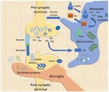
          The synaptic cleft has been vastly investigated in the last decades, leading to a novel and fascinating model of the functional and structural modifications linked to synaptic transmission and brain processing. The classic neurocentric model encompassing the neuronal pre- and post-synaptic terminals partly explains the fine-tuned plastic modifications under both pathological and physiological circumstances. Recent experimental evidence has incontrovertibly added oligodendrocytes, astrocytes, and microglia as pivotal elements for synapse formation and remodeling (tripartite synapse) in both the developing and adult brain. Moreover, synaptic plasticity and its pathological counterpart (maladaptive plasticity) have shown a deep connection with other molecular elements of the extracellular matrix (ECM), once considered as a mere extracellular structural scaffold altogether with the cellular glue (i.e., glia). The ECM adds another level of complexity to the modern model of the synapse, particularly, for the long-term plasticity and circuit maintenance. This model, called tetrapartite synapse, can be further implemented by including the neurovascular unit (NVU) and the immune system. Although they were considered so far as tightly separated from the central nervous system (CNS) plasticity, at least in physiological conditions, recent evidence endorsed these elements as structural and paramount actors in synaptic plasticity. This scenario is, as far as speculations and evidence have shown, a consistent model for both adaptive and maladaptive plasticity. However, a comprehensive understanding of brain processes and circuitry complexity is still lacking. Here we propose that a better interpretation of the CNS complexity can be granted by a systems biology approach through the construction of predictive molecular models that enable to enlighten the regulatory logic of the complex molecular networks underlying brain function in health and disease, thus opening the way to more effective treatments.

          Related collections

          Most cited references135

          • Record: found
          • Abstract: found
          • Article: not found

          Uniquely hominid features of adult human astrocytes.

          Defining the microanatomic differences between the human brain and that of other mammals is key to understanding its unique computational power. Although much effort has been devoted to comparative studies of neurons, astrocytes have received far less attention. We report here that protoplasmic astrocytes in human neocortex are 2.6-fold larger in diameter and extend 10-fold more GFAP (glial fibrillary acidic protein)-positive primary processes than their rodent counterparts. In cortical slices prepared from acutely resected surgical tissue, protoplasmic astrocytes propagate Ca(2+) waves with a speed of 36 microm/s, approximately fourfold faster than rodent. Human astrocytes also transiently increase cystosolic Ca(2+) in response to glutamatergic and purinergic receptor agonists. The human neocortex also harbors several anatomically defined subclasses of astrocytes not represented in rodents. These include a population of astrocytes that reside in layers 5-6 and extend long fibers characterized by regularly spaced varicosities. Another specialized type of astrocyte, the interlaminar astrocyte, abundantly populates the superficial cortical layers and extends long processes without varicosities to cortical layers 3 and 4. Human fibrous astrocytes resemble their rodent counterpart but are larger in diameter. Thus, human cortical astrocytes are both larger, and structurally both more complex and more diverse, than those of rodents. On this basis, we posit that this astrocytic complexity has permitted the increased functional competence of the adult human brain.
            Bookmark
            • Record: found
            • Abstract: found
            • Article: not found

            Tripartite synapses: glia, the unacknowledged partner.

            According to the classical view of the nervous system, the numerically superior glial cells have inferior roles in that they provide an ideal environment for neuronal-cell function. However, there is a wave of new information suggesting that glia are intimately involved in the active control of neuronal activity and synaptic neurotransmission. Recent evidence shows that glia respond to neuronal activity with an elevation of their internal Ca2+ concentration, which triggers the release of chemical transmitters from glia themselves and, in turn, causes feedback regulation of neuronal activity and synaptic strength. In view of these new insights, this article suggests that perisynaptic Schwann cells and synaptically associated astrocytes should be viewed as integral modulatory elements of tripartite synapses.
              Bookmark
              • Record: found
              • Abstract: found
              • Article: not found

              Brain-wide pathway for waste clearance captured by contrast-enhanced MRI.

              The glymphatic system is a recently defined brain-wide paravascular pathway for cerebrospinal fluid (CSF) and interstitial fluid (ISF) exchange that facilitates efficient clearance of solutes and waste from the brain. CSF enters the brain along para-arterial channels to exchange with ISF, which is in turn cleared from the brain along para-venous pathways. Because soluble amyloid β clearance depends on glymphatic pathway function, we proposed that failure of this clearance system contributes to amyloid plaque deposition and Alzheimer's disease progression. Here we provide proof of concept that glymphatic pathway function can be measured using a clinically relevant imaging technique. Dynamic contrast-enhanced MRI was used to visualize CSF-ISF exchange across the rat brain following intrathecal paramagnetic contrast agent administration. Key features of glymphatic pathway function were confirmed, including visualization of para-arterial CSF influx and molecular size-dependent CSF-ISF exchange. Whole-brain imaging allowed the identification of two key influx nodes at the pituitary and pineal gland recesses, while dynamic MRI permitted the definition of simple kinetic parameters to characterize glymphatic CSF-ISF exchange and solute clearance from the brain. We propose that this MRI approach may provide the basis for a wholly new strategy to evaluate Alzheimer's disease susceptibility and progression in the live human brain.
                Bookmark

                Author and article information

                Journal
                Int J Mol Sci
                Int J Mol Sci
                ijms
                International Journal of Molecular Sciences
                MDPI
                1422-0067
                24 February 2020
                February 2020
                : 21
                : 4
                : 1539
                Affiliations
                [1 ]Laboratory of Morphology of Neuronal Network, Department of Public Medicine, University of Campania “Luigi Vanvitelli”, 80138 Napoli, Italy; delucaciro88@ 123456gmail.com (C.D.L.); assunta-1989@ 123456hotmail.it (A.V.); michele.papa@ 123456unicampania.it (M.P.)
                [2 ]Laboratory of Neuroscience “R. Levi-Montalcini”, Dept. of Biotechnology and Biosciences, University of Milano-Bicocca, 20126 Milano, Italy
                [3 ]SYSBIO Centre of Systems Biology ISBE.ITALY, University of Milano-Bicocca, 20126 Milano, Italy; lilia.alberghina@ 123456gmail.com
                Author notes
                Author information
                https://orcid.org/0000-0002-7971-4289
                https://orcid.org/0000-0002-6609-7453
                Article
                ijms-21-01539
                10.3390/ijms21041539
                7073232
                32102370
                6c2c7064-b3db-4525-ad7b-efee438da1cd
                © 2020 by the authors.

                Licensee MDPI, Basel, Switzerland. This article is an open access article distributed under the terms and conditions of the Creative Commons Attribution (CC BY) license ( http://creativecommons.org/licenses/by/4.0/).

                History
                : 31 December 2019
                : 20 February 2020
                Categories
                Review

                Molecular biology
                glia,tripartite synapse,synaptic plasticity,neurovascular unit,systems biology
                Molecular biology
                glia, tripartite synapse, synaptic plasticity, neurovascular unit, systems biology

                Comments

                Comment on this article