4
views
0
recommends
+1 Recommend
0 collections
    0
    shares
      • Record: found
      • Abstract: found
      • Article: found
      Is Open Access

      Monitoring the maturation of the sarcomere network: a super-resolution microscopy-based approach

      research-article

      Read this article at

      Bookmark
          There is no author summary for this article yet. Authors can add summaries to their articles on ScienceOpen to make them more accessible to a non-specialist audience.

          Abstract

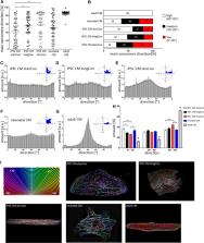
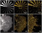
          The in vitro generation of human cardiomyocytes derived from induced pluripotent stem cells (iPSC) is of great importance for cardiac disease modeling, drug-testing applications and for regenerative medicine. Despite the development of various cultivation strategies, a sufficiently high degree of maturation is still a decisive limiting factor for the successful application of these cardiac cells. The maturation process includes, among others, the proper formation of sarcomere structures, mediating the contraction of cardiomyocytes. To precisely monitor the maturation of the contractile machinery, we have established an imaging-based strategy that allows quantitative evaluation of important parameters, defining the quality of the sarcomere network. iPSC-derived cardiomyocytes were subjected to different culture conditions to improve sarcomere formation, including prolonged cultivation time and micro patterned surfaces. Fluorescent images of α-actinin were acquired using super-resolution microscopy. Subsequently, we determined cell morphology, sarcomere density, filament alignment, z-Disc thickness and sarcomere length of iPSC-derived cardiomyocytes. Cells from adult and neonatal heart tissue served as control. Our image analysis revealed a profound effect on sarcomere content and filament orientation when iPSC-derived cardiomyocytes were cultured on structured, line-shaped surfaces. Similarly, prolonged cultivation time had a beneficial effect on the structural maturation, leading to a more adult-like phenotype. Automatic evaluation of the sarcomere filaments by machine learning validated our data. Moreover, we successfully transferred this approach to skeletal muscle cells, showing an improved sarcomere formation cells over different differentiation periods. Overall, our image-based workflow can be used as a straight-forward tool to quantitatively estimate the structural maturation of contractile cells. As such, it can support the establishment of novel differentiation protocols to enhance sarcomere formation and maturity.

          Supplementary Information

          The online version contains supplementary material available at 10.1007/s00018-022-04196-3.

          Related collections

          Most cited references64

          • Record: found
          • Abstract: found
          • Article: not found

          Induction of pluripotent stem cells from mouse embryonic and adult fibroblast cultures by defined factors.

          Differentiated cells can be reprogrammed to an embryonic-like state by transfer of nuclear contents into oocytes or by fusion with embryonic stem (ES) cells. Little is known about factors that induce this reprogramming. Here, we demonstrate induction of pluripotent stem cells from mouse embryonic or adult fibroblasts by introducing four factors, Oct3/4, Sox2, c-Myc, and Klf4, under ES cell culture conditions. Unexpectedly, Nanog was dispensable. These cells, which we designated iPS (induced pluripotent stem) cells, exhibit the morphology and growth properties of ES cells and express ES cell marker genes. Subcutaneous transplantation of iPS cells into nude mice resulted in tumors containing a variety of tissues from all three germ layers. Following injection into blastocysts, iPS cells contributed to mouse embryonic development. These data demonstrate that pluripotent stem cells can be directly generated from fibroblast cultures by the addition of only a few defined factors.
            Bookmark
            • Record: found
            • Abstract: found
            • Article: found
            Is Open Access

            ThunderSTORM: a comprehensive ImageJ plug-in for PALM and STORM data analysis and super-resolution imaging

            Summary: ThunderSTORM is an open-source, interactive and modular plug-in for ImageJ designed for automated processing, analysis and visualization of data acquired by single-molecule localization microscopy methods such as photo-activated localization microscopy and stochastic optical reconstruction microscopy. ThunderSTORM offers an extensive collection of processing and post-processing methods so that users can easily adapt the process of analysis to their data. ThunderSTORM also offers a set of tools for creation of simulated data and quantitative performance evaluation of localization algorithms using Monte Carlo simulations. Availability and implementation: ThunderSTORM and the online documentation are both freely accessible at https://code.google.com/p/thunder-storm/ Contact: guy.hagen@lf1.cuni.cz Supplementary information: Supplementary data are available at Bioinformatics online.
              Bookmark
              • Record: found
              • Abstract: found
              • Article: not found

              Experimental investigation of collagen waviness and orientation in the arterial adventitia using confocal laser scanning microscopy.

              Mechanical properties of the adventitia are largely determined by the organization of collagen fibers. Measurements on the waviness and orientation of collagen, particularly at the zero-stress state, are necessary to relate the structural organization of collagen to the mechanical response of the adventitia. Using the fluorescence collagen marker CNA38-OG488 and confocal laser scanning microscopy, we imaged collagen fibers in the adventitia of rabbit common carotid arteries ex vivo. The arteries were cut open along their longitudinal axes to get the zero-stress state. We used semi-manual and automatic techniques to measure parameters related to the waviness and orientation of fibers. Our results showed that the straightness parameter (defined as the ratio between the distances of endpoints of a fiber to its length) was distributed with a beta distribution (mean value 0.72, variance 0.028) and did not depend on the mean angle orientation of fibers. Local angular density distributions revealed four axially symmetric families of fibers with mean directions of 0°, 90°, 43° and -43°, with respect to the axial direction of the artery, and corresponding circular standard deviations of 40°, 47°, 37° and 37°. The distribution of local orientations was shifted to the circumferential direction when measured in arteries at the zero-load state (intact), as compared to arteries at the zero-stress state (cut-open). Information on collagen fiber waviness and orientation, such as obtained in this study, could be used to develop structural models of the adventitia, providing better means for analyzing and understanding the mechanical properties of vascular wall.
                Bookmark

                Author and article information

                Contributors
                robert.david@med.uni-rostock.de
                Journal
                Cell Mol Life Sci
                Cell Mol Life Sci
                Cellular and Molecular Life Sciences
                Springer International Publishing (Cham )
                1420-682X
                1420-9071
                23 February 2022
                23 February 2022
                2022
                : 79
                : 3
                : 149
                Affiliations
                [1 ]GRID grid.413108.f, ISNI 0000 0000 9737 0454, Department of Cardiac Surgery, Reference and Translation Center for Cardiac Stem Cell Therapy (RTC), , Rostock University Medical Center, ; 18057 Rostock, Germany
                [2 ]GRID grid.10493.3f, ISNI 0000000121858338, Faculty of Interdisciplinary Research, Department Life, Light and Matter, , University Rostock, ; 18059 Rostock, Germany
                [3 ]GRID grid.10493.3f, ISNI 0000000121858338, Department of Systems Biology and Bioinformatics, , University of Rostock, ; Rostock, Germany
                [4 ]GRID grid.413108.f, ISNI 0000 0000 9737 0454, Research Laboratory for Biomechanics and Implant Technology, Department of Orthopedics, , Rostock University Medical Center, ; 18057 Rostock, Germany
                [5 ]GRID grid.10493.3f, ISNI 0000000121858338, Department of Operative Dentistry and Periodontology, , Rostock University Medical Centre, ; 18057 Rostock, Germany
                [6 ]GRID grid.11956.3a, ISNI 0000 0001 2214 904X, Stellenbosch Institute of Advanced Study (STIAS), , Wallenberg Research Centre at Stellenbosch University, ; Stellenbosch, 7602 South Africa
                Author information
                http://orcid.org/0000-0003-2821-2323
                Article
                4196
                10.1007/s00018-022-04196-3
                8866374
                35199227
                6c7186f2-5ba6-493f-ba2c-0c67e7c24717
                © The Author(s) 2022

                Open AccessThis article is licensed under a Creative Commons Attribution 4.0 International License, which permits use, sharing, adaptation, distribution and reproduction in any medium or format, as long as you give appropriate credit to the original author(s) and the source, provide a link to the Creative Commons licence, and indicate if changes were made. The images or other third party material in this article are included in the article's Creative Commons licence, unless indicated otherwise in a credit line to the material. If material is not included in the article's Creative Commons licence and your intended use is not permitted by statutory regulation or exceeds the permitted use, you will need to obtain permission directly from the copyright holder. To view a copy of this licence, visit http://creativecommons.org/licenses/by/4.0/.

                History
                : 18 August 2021
                : 22 January 2022
                : 5 February 2022
                Funding
                Funded by: EU Structural Fund
                Award ID: ESF/14-BM-A55-0024/18
                Funded by: Josef and Käthe Klinz foundation
                Award ID: T319/29737/2017
                Award Recipient :
                Funded by: FORUN Program University Medical Center Rostock
                Award ID: 889003
                Award ID: 889001
                Award Recipient :
                Funded by: FundRef http://dx.doi.org/10.13039/501100001659, Deutsche Forschungsgemeinschaft;
                Award ID: DA1296/6-1
                Award Recipient :
                Funded by: FundRef http://dx.doi.org/10.13039/501100002347, Bundesministerium für Bildung und Forschung;
                Award ID: VIP+ 00240
                Award Recipient :
                Funded by: FundRef http://dx.doi.org/10.13039/501100005971, Deutsche Herzstiftung;
                Award ID: F/01/12
                Award Recipient :
                Funded by: DAMP Foundation
                Funded by: Universitätsmedizin Rostock (8980)
                Categories
                Original Article
                Custom metadata
                © Springer Nature Switzerland AG 2022

                Molecular biology
                ipsc cardiomyocytes,sarcomere network,maturation,super resolution microscopy
                Molecular biology
                ipsc cardiomyocytes, sarcomere network, maturation, super resolution microscopy

                Comments

                Comment on this article