5
views
0
recommends
+1 Recommend
0 collections
    0
    shares
      • Record: found
      • Abstract: found
      • Article: found
      Is Open Access

      The Influence of Aging, Hearing, and Tinnitus on the Morphology of Cortical Gray Matter, Amygdala, and Hippocampus

      research-article

      Read this article at

      Bookmark
          There is no author summary for this article yet. Authors can add summaries to their articles on ScienceOpen to make them more accessible to a non-specialist audience.

          Abstract

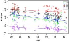
          Age related hearing loss (presbycusis) is a natural process represented by elevated auditory thresholds and decreased speech intelligibility, especially in noisy conditions. Tinnitus is a phantom sound that also potentially leads to cortical changes, with its highest occurrence coinciding with the clinical onset of presbycusis. The aim of our project was to identify age, hearing loss and tinnitus related structural changes, within the auditory system and associated structures. Groups of subjects with presbycusis and tinnitus (22 subjects), with only presbycusis (24 subjects), young tinnitus patients with normal hearing (10 subjects) and young controls (17 subjects), underwent an audiological examination to characterize hearing loss and tinnitus. In addition, MRI (3T MR system, analysis in Freesurfer software) scans were used to identify changes in the cortical and subcortical structures. The following areas of the brain were analyzed: Heschl gyrus (HG), planum temporale (PT), primary visual cortex (V1), gyrus parahippocampus (PH), anterior insula (Ins), amygdala (Amg), and hippocampus (HP). A statistical analysis was performed in R framework using linear mixed-effects models with explanatory variables: age, tinnitus, laterality and hearing. In all of the cortical structures, the gray matter thickness decreased significantly with aging without having an effect on laterality (differences between the left and right hemispheres). The decrease in the gray matter thickness was faster in the HG, PT and Ins in comparison with the PH and V1. Aging did not influence the surface of the cortical areas, however there were differences between the surface size of the reported regions in the left and right hemispheres. Hearing loss caused only a borderline decrease of the cortical surface in the HG. Tinnitus was accompanied by a borderline decrease of the Ins surface and led to an increase in the volume of Amy and HP. In summary, aging is accompanied by a decrease in the cortical gray matter thickness; hearing loss only has a limited effect on the structure of the investigated cortical areas and tinnitus causes structural changes which are predominantly within the limbic system and insula, with the structure of the auditory system only being minimally affected.

          Related collections

          Most cited references108

          • Record: found
          • Abstract: found
          • Article: not found

          Cortical surface-based analysis. I. Segmentation and surface reconstruction.

          Several properties of the cerebral cortex, including its columnar and laminar organization, as well as the topographic organization of cortical areas, can only be properly understood in the context of the intrinsic two-dimensional structure of the cortical surface. In order to study such cortical properties in humans, it is necessary to obtain an accurate and explicit representation of the cortical surface in individual subjects. Here we describe a set of automated procedures for obtaining accurate reconstructions of the cortical surface, which have been applied to data from more than 100 subjects, requiring little or no manual intervention. Automated routines for unfolding and flattening the cortical surface are described in a companion paper. These procedures allow for the routine use of cortical surface-based analysis and visualization methods in functional brain imaging. Copyright 1999 Academic Press.
            Bookmark
            • Record: found
            • Abstract: found
            • Article: not found

            The minimal preprocessing pipelines for the Human Connectome Project.

            The Human Connectome Project (HCP) faces the challenging task of bringing multiple magnetic resonance imaging (MRI) modalities together in a common automated preprocessing framework across a large cohort of subjects. The MRI data acquired by the HCP differ in many ways from data acquired on conventional 3 Tesla scanners and often require newly developed preprocessing methods. We describe the minimal preprocessing pipelines for structural, functional, and diffusion MRI that were developed by the HCP to accomplish many low level tasks, including spatial artifact/distortion removal, surface generation, cross-modal registration, and alignment to standard space. These pipelines are specially designed to capitalize on the high quality data offered by the HCP. The final standard space makes use of a recently introduced CIFTI file format and the associated grayordinate spatial coordinate system. This allows for combined cortical surface and subcortical volume analyses while reducing the storage and processing requirements for high spatial and temporal resolution data. Here, we provide the minimum image acquisition requirements for the HCP minimal preprocessing pipelines and additional advice for investigators interested in replicating the HCP's acquisition protocols or using these pipelines. Finally, we discuss some potential future improvements to the pipelines. Copyright © 2013 Elsevier Inc. All rights reserved.
              Bookmark
              • Record: found
              • Abstract: found
              • Article: not found

              Dissociable intrinsic connectivity networks for salience processing and executive control.

              Variations in neural circuitry, inherited or acquired, may underlie important individual differences in thought, feeling, and action patterns. Here, we used task-free connectivity analyses to isolate and characterize two distinct networks typically coactivated during functional MRI tasks. We identified a "salience network," anchored by dorsal anterior cingulate (dACC) and orbital frontoinsular cortices with robust connectivity to subcortical and limbic structures, and an "executive-control network" that links dorsolateral frontal and parietal neocortices. These intrinsic connectivity networks showed dissociable correlations with functions measured outside the scanner. Prescan anxiety ratings correlated with intrinsic functional connectivity of the dACC node of the salience network, but with no region in the executive-control network, whereas executive task performance correlated with lateral parietal nodes of the executive-control network, but with no region in the salience network. Our findings suggest that task-free analysis of intrinsic connectivity networks may help elucidate the neural architectures that support fundamental aspects of human behavior.
                Bookmark

                Author and article information

                Contributors
                Journal
                Front Aging Neurosci
                Front Aging Neurosci
                Front. Aging Neurosci.
                Frontiers in Aging Neuroscience
                Frontiers Media S.A.
                1663-4365
                04 December 2020
                2020
                : 12
                : 553461
                Affiliations
                [1] 1Department of Auditory Neuroscience, Institute of Experimental Medicine of the Czech Academy of Sciences , Prague, Czechia
                [2] 2Department of Otorhinolaryngology, 3rd Faculty of Medicine, Faculty Hospital Kralovske Vinohrady, Charles University , Prague, Czechia
                [3] 3MR Unit, Institute of Clinical and Experimental Medicine , Prague, Czechia
                [4] 4Department of Otorhinolaryngology and Head and Neck Surgery, 1st Faculty of Medicine, University Hospital Motol, Charles University , Prague, Czechia
                Author notes

                Edited by: Tobias Kleinjung, University of Zurich, Switzerland

                Reviewed by: Benjamin Joseph Zimmerman, University of Illinois at Urbana-Champaign, United States; Cris Lanting, Radboud University Nijmegen Medical Centre, Netherlands

                Article
                10.3389/fnagi.2020.553461
                7746808
                33343328
                6eaae93a-0125-4c1c-83d9-924f15c1bd12
                Copyright © 2020 Profant, Škoch, Tintěra, Svobodová, Kuchárová, Svobodová Burianová and Syka.

                This is an open-access article distributed under the terms of the Creative Commons Attribution License (CC BY). The use, distribution or reproduction in other forums is permitted, provided the original author(s) and the copyright owner(s) are credited and that the original publication in this journal is cited, in accordance with accepted academic practice. No use, distribution or reproduction is permitted which does not comply with these terms.

                History
                : 18 April 2020
                : 12 November 2020
                Page count
                Figures: 7, Tables: 1, Equations: 0, References: 109, Pages: 16, Words: 11965
                Funding
                Funded by: Grantová Agentura Ceské Republiky 10.13039/501100001824
                Categories
                Neuroscience
                Original Research

                Neurosciences
                presbycusis,tinnitus,auditory system,limbic system,morphometry
                Neurosciences
                presbycusis, tinnitus, auditory system, limbic system, morphometry

                Comments

                Comment on this article