20
views
0
recommends
+1 Recommend
0 collections
    0
    shares
      • Record: found
      • Abstract: found
      • Article: found
      Is Open Access

      Reduced TRMU expression increases the sensitivity of hair-cell-like HEI-OC-1 cells to neomycin damage in vitro

      research-article

      Read this article at

      Bookmark
          There is no author summary for this article yet. Authors can add summaries to their articles on ScienceOpen to make them more accessible to a non-specialist audience.

          Abstract

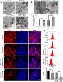
          Aminoglycosides are ototoxic to the cochlear hair cells, and mitochondrial dysfunction is one of the major mechanisms behind ototoxic drug-induced hair cell death. TRMU (tRNA 5-methylaminomethyl-2-thiouridylate methyltransferase) is a mitochondrial protein that participates in mitochondrial tRNA modifications, but the role of TRMU in aminoglycoside-induced ototoxicity remains to be elucidated. In this study, we took advantage of the HEI-OC-1 cell line to investigate the role of TRMU in aminoglycoside-induced cell death. We found that TRMU is expressed in both hair cells and HEI-OC-1 cells, and its expression is significantly decreased after 24 h neomycin treatment. We then downregulated TRMU expression with siRNA and found that cell death and apoptosis were significantly increased after neomycin injury. Furthermore, when we down-regulated TRMU expression, we observed significantly increased mitochondrial dysfunction and increased levels of reactive oxygen species (ROS) after neomycin injury, suggesting that TRMU regulates mitochondrial function and ROS levels. Lastly, the antioxidant N-acetylcysteine rescued the mitochondrial dysfunction and cell apoptosis that was induced by TRMU downregulation, suggesting that ROS accumulation contributed to the increased aminoglycosides sensitivity of HEI-OC-1 cells after TRMU downregulation. This study provides evidence that TRMU might be a new therapeutic target for the prevention of aminoglycoside-induced hair cell death.

          Related collections

          Most cited references46

          • Record: found
          • Abstract: found
          • Article: not found

          Mitochondrial transcription factor A regulates mtDNA copy number in mammals.

          Mitochondrial DNA (mtDNA) copy number regulation is altered in several human mtDNA-mutation diseases and it is also important in a variety of normal physiological processes. Mitochondrial transcription factor A (TFAM) is essential for human mtDNA transcription and we demonstrate here that it is also a key regulator of mtDNA copy number. We initially performed in vitro transcription studies and determined that the human TFAM protein is a poor activator of mouse mtDNA transcription, despite its high capacity for unspecific DNA binding. Next, we generated P1 artificial chromosome (PAC) transgenic mice ubiquitously expressing human TFAM. The introduced human TFAM gene was regulated in a similar fashion as the endogenous mouse Tfam gene and expression of the human TFAM protein in the mouse did not result in down-regulation of the endogenous expression. The PAC-TFAM mice thus had a net overexpression of TFAM protein and this resulted in a general increase of mtDNA copy number. We used a combination of mice with TFAM overexpression and TFAM knockout and demonstrated that mtDNA copy number is directly proportional to the total TFAM protein levels also in mouse embryos. Interestingly, the expression of human TFAM in the mouse results in up-regulation of mtDNA copy number without increasing respiratory chain capacity or mitochondrial mass. It is thus possible to experimentally dissociate mtDNA copy number regulation from mtDNA expression and mitochondrial biogenesis in mammals in vivo. In conclusion, our results provide genetic evidence for a novel role for TFAM in direct regulation of mtDNA copy number in mammals.
            Bookmark
            • Record: found
            • Abstract: found
            • Article: not found

            Essential role of the mitochondrial apoptosis-inducing factor in programmed cell death.

            Programmed cell death is a fundamental requirement for embryogenesis, organ metamorphosis and tissue homeostasis. In mammals, release of mitochondrial cytochrome c leads to the cytosolic assembly of the apoptosome-a caspase activation complex involving Apaf1 and caspase-9 that induces hallmarks of apoptosis. There are, however, mitochondrially regulated cell death pathways that are independent of Apaf1/caspase-9. We have previously cloned a molecule associated with programmed cell death called apoptosis-inducing factor (AIF). Like cytochrome c, AIF is localized to mitochondria and released in response to death stimuli. Here we show that genetic inactivation of AIF renders embryonic stem cells resistant to cell death after serum deprivation. Moreover, AIF is essential for programmed cell death during cavitation of embryoid bodies-the very first wave of cell death indispensable for mouse morphogenesis. AIF-dependent cell death displays structural features of apoptosis, and can be genetically uncoupled from Apaf1 and caspase-9 expression. Our data provide genetic evidence for a caspase-independent pathway of programmed cell death that controls early morphogenesis.
              Bookmark
              • Record: found
              • Abstract: found
              • Article: not found

              Use of whole-exome sequencing to determine the genetic basis of multiple mitochondrial respiratory chain complex deficiencies.

              Mitochondrial disorders have emerged as a common cause of inherited disease, but their diagnosis remains challenging. Multiple respiratory chain complex defects are particularly difficult to diagnose at the molecular level because of the massive number of nuclear genes potentially involved in intramitochondrial protein synthesis, with many not yet linked to human disease. To determine the molecular basis of multiple respiratory chain complex deficiencies. We studied 53 patients referred to 2 national centers in the United Kingdom and Germany between 2005 and 2012. All had biochemical evidence of multiple respiratory chain complex defects but no primary pathogenic mitochondrial DNA mutation. Whole-exome sequencing was performed using 62-Mb exome enrichment, followed by variant prioritization using bioinformatic prediction tools, variant validation by Sanger sequencing, and segregation of the variant with the disease phenotype in the family. Presumptive causal variants were identified in 28 patients (53%; 95% CI, 39%-67%) and possible causal variants were identified in 4 (8%; 95% CI, 2%-18%). Together these accounted for 32 patients (60% 95% CI, 46%-74%) and involved 18 different genes. These included recurrent mutations in RMND1, AARS2, and MTO1, each on a haplotype background consistent with a shared founder allele, and potential novel mutations in 4 possible mitochondrial disease genes (VARS2, GARS, FLAD1, and PTCD1). Distinguishing clinical features included deafness and renal involvement associated with RMND1 and cardiomyopathy with AARS2 and MTO1. However, atypical clinical features were present in some patients, including normal liver function and Leigh syndrome (subacute necrotizing encephalomyelopathy) seen in association with TRMU mutations and no cardiomyopathy with founder SCO2 mutations. It was not possible to confidently identify the underlying genetic basis in 21 patients (40%; 95% CI, 26%-54%). Exome sequencing enhances the ability to identify potential nuclear gene mutations in patients with biochemically defined defects affecting multiple mitochondrial respiratory chain complexes. Additional study is required in independent patient populations to determine the utility of this approach in comparison with traditional diagnostic methods.
                Bookmark

                Author and article information

                Journal
                Sci Rep
                Sci Rep
                Scientific Reports
                Nature Publishing Group
                2045-2322
                13 July 2016
                2016
                : 6
                : 29621
                Affiliations
                [1 ]State Key Laboratory of Bioelectronics, Institute of Life Sciences, Southeast University , Nanjing 210096, China
                [2 ]Department of Otorhinolaryngology, Hearing Research Institute, Affiliated Eye and ENT Hospital of Fudan University , Shanghai 200031, China
                [3 ]MOE Key Laboratory of Developmental Genes and Human Disease, Institute of Life Sciences, Southeast University , Nanjing 210096, China
                [4 ]Co-Innovation Center of Neuroregeneration, Nantong University , Nantong 226001, China
                [5 ]Department of Otolaryngology, Affiliated Drum Tower Hospital of Nanjing University Medical School , Nanjing 210008, China
                [6 ]Medical School, Southeast University , Nanjing 210096, China
                [7 ]Institutes of Life Sciences, Fudan University , Shanghai 200032, China
                Author notes
                [*]

                These authors contributed equally to this work.

                Article
                srep29621
                10.1038/srep29621
                4942793
                27405449
                6f9278ff-f8bb-4ee5-ab95-f15d9f80ac76
                Copyright © 2016, Macmillan Publishers Limited

                This work is licensed under a Creative Commons Attribution 4.0 International License. The images or other third party material in this article are included in the article’s Creative Commons license, unless indicated otherwise in the credit line; if the material is not included under the Creative Commons license, users will need to obtain permission from the license holder to reproduce the material. To view a copy of this license, visit http://creativecommons.org/licenses/by/4.0/

                History
                : 04 April 2016
                : 20 June 2016
                Categories
                Article

                Uncategorized
                Uncategorized

                Comments

                Comment on this article