17
views
0
recommends
+1 Recommend
0 collections
    4
    shares
      • Record: found
      • Abstract: found
      • Article: found
      Is Open Access

      STED nanoscopy with fluorescent quantum dots

      research-article

      Read this article at

      Bookmark
          There is no author summary for this article yet. Authors can add summaries to their articles on ScienceOpen to make them more accessible to a non-specialist audience.

          Abstract

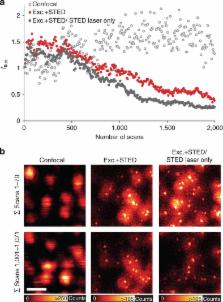
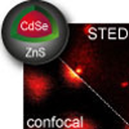
          The widely popular class of quantum-dot molecular labels could so far not be utilized as standard fluorescent probes in STED (stimulated emission depletion) nanoscopy. This is because broad quantum-dot excitation spectra extend deeply into the spectral bands used for STED, thus compromising the transient fluorescence silencing required for attaining super-resolution. Here we report the discovery that STED nanoscopy of several red-emitting commercially available quantum dots is in fact successfully realized by the increasingly popular 775 nm STED laser light. A resolution of presently ∼50 nm is demonstrated for single quantum dots, and sub-diffraction resolution is further shown for imaging of quantum-dot-labelled vimentin filaments in fibroblasts. The high quantum-dot photostability enables repeated STED recordings with >1,000 frames. In addition, we have evidence that the tendency of quantum-dot labels to blink is largely suppressed by combined action of excitation and STED beams. Quantum-dot STED significantly expands the realm of application of STED nanoscopy, and, given the high stability of these probes, holds promise for extended time-lapse imaging.

          Abstract

          STED nanoscopy enables sub-diffraction imaging with a wide range of fluorescent probes. Here, the authors show that a bright and very photostable class of fluorescent quantum dots can be super-resolved with STED as biolabels in cellular contexts.

          Related collections

          Most cited references13

          • Record: found
          • Abstract: found
          • Article: not found

          Quantum dot bioconjugates for ultrasensitive nonisotopic detection.

          W Chan, S Nie (1998)
          Highly luminescent semiconductor quantum dots (zinc sulfide-capped cadmium selenide) have been covalently coupled to biomolecules for use in ultrasensitive biological detection. In comparison with organic dyes such as rhodamine, this class of luminescent labels is 20 times as bright, 100 times as stable against photobleaching, and one-third as wide in spectral linewidth. These nanometer-sized conjugates are water-soluble and biocompatible. Quantum dots that were labeled with the protein transferrin underwent receptor-mediated endocytosis in cultured HeLa cells, and those dots that were labeled with immunomolecules recognized specific antibodies or antigens.
            Bookmark
            • Record: found
            • Abstract: found
            • Article: not found

            Far-field optical nanoscopy.

            In 1873, Ernst Abbe discovered what was to become a well-known paradigm: the inability of a lens-based optical microscope to discern details that are closer together than half of the wavelength of light. However, for its most popular imaging mode, fluorescence microscopy, the diffraction barrier is crumbling. Here, I discuss the physical concepts that have pushed fluorescence microscopy to the nanoscale, once the prerogative of electron and scanning probe microscopes. Initial applications indicate that emergent far-field optical nanoscopy will have a strong impact in the life sciences and in other areas benefiting from nanoscale visualization.
              Bookmark
              • Record: found
              • Abstract: found
              • Article: not found

              Long-term multiple color imaging of live cells using quantum dot bioconjugates.

              Luminescent quantum dots (QDs)--semiconductor nanocrystals--are a promising alternative to organic dyes for fluorescence-based applications. We have developed procedures for using QDs to label live cells and have demonstrated their use for long-term multicolor imaging of live cells. The two approaches presented are (i) endocytic uptake of QDs and (ii) selective labeling of cell surface proteins with QDs conjugated to antibodies. Live cells labeled using these approaches were used for long-term multicolor imaging. The cells remained stably labeled for over a week as they grew and developed. These approaches should permit the simultaneous study of multiple cells over long periods of time as they proceed through growth and development.
                Bookmark

                Author and article information

                Journal
                Nat Commun
                Nat Commun
                Nature Communications
                Nature Pub. Group
                2041-1723
                18 May 2015
                2015
                : 6
                : 7127
                Affiliations
                [1 ]German Cancer Research Center (DKFZ), Optical Nanoscopy Division , Im Neuenheimer Feld 280, Heidelberg 69120, Germany
                [2 ]Department of NanoBiophotonics, Max Planck Institute for Biophysical Chemistry , Am Fassberg 11, Göttingen 37077, Germany
                Author notes
                [*]

                These authors contributed equally to this work

                Author information
                http://orcid.org/0000-0003-3159-1416
                Article
                ncomms8127
                10.1038/ncomms8127
                4479004
                25980788
                702f0b1e-b67a-4b15-a1d1-34a947d1d5a9
                Copyright © 2015, Nature Publishing Group, a division of Macmillan Publishers Limited. All Rights Reserved.

                This work is licensed under a Creative Commons Attribution 4.0 International License. The images or other third party material in this article are included in the article's Creative Commons license, unless indicated otherwise in the credit line; if the material is not included under the Creative Commons license, users will need to obtain permission from the license holder to reproduce the material. To view a copy of this license, visit http://creativecommons.org/licenses/by/4.0/

                History
                : 17 January 2015
                : 02 April 2015
                Categories
                Article

                Uncategorized
                Uncategorized

                Comments

                Comment on this article