3
views
0
recommends
+1 Recommend
0 collections
    0
    shares
      • Record: found
      • Abstract: found
      • Article: found
      Is Open Access

      RGC-specific ATF4 and/or CHOP deletion rescues glaucomatous neurodegeneration and visual function

      research-article

      Read this article at

      Bookmark
          There is no author summary for this article yet. Authors can add summaries to their articles on ScienceOpen to make them more accessible to a non-specialist audience.

          Abstract

          Endoplasmic reticulum (ER) stress has been linked with various acute and chronic neurodegenerative diseases. We previously found that optic nerve (ON) injury and diseases induce neuronal ER stress in retinal ganglion cells (RGCs). We further demonstrated that germline deletion of CHOP preserves the structure and function of both RGC somata and axons in mouse glaucoma models. Here we report that RGC-specific deletion of CHOP and/or its upstream regulator ATF4 synergistically promotes RGC and ON survival and preserves visual function in mouse ON crush and silicone oil-induced ocular hypertension (SOHU) glaucoma models. Consistently, topical application of the ATF4/CHOP chemical inhibitor ISRIB or RGC-specific CRISPR-mediated knockdown of the ATF4 downstream effector Gadd45a also delivers significant neuroprotection in the SOHU glaucoma model. These studies suggest that blocking the neuronal intrinsic ATF4/CHOP axis of ER stress is a promising neuroprotection strategy for neurodegeneration.
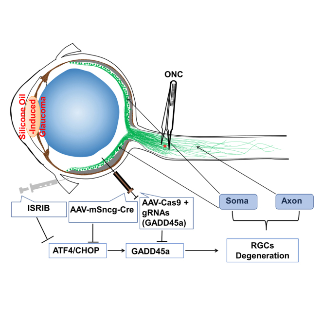
          Graphical abstract

          Abstract

          Hu and colleagues present experimental evidence that neuronal intrinsic blocking of ATF4 and/or CHOP, ATF4 downstream molecule, Gadd45a, and local delivery of an ATF4/CHOP inhibitor, ISRIB, prevent glaucomatous neurodegeneration, indicating a promising neuroprotective gene therapy strategy for retinal ganglion cells in traumatic and glaucomatous optic neuropathies.

          Related collections

          Most cited references76

          • Record: found
          • Abstract: found
          • Article: not found

          The unfolded protein response: from stress pathway to homeostatic regulation.

          The vast majority of proteins that a cell secretes or displays on its surface first enter the endoplasmic reticulum (ER), where they fold and assemble. Only properly assembled proteins advance from the ER to the cell surface. To ascertain fidelity in protein folding, cells regulate the protein-folding capacity in the ER according to need. The ER responds to the burden of unfolded proteins in its lumen (ER stress) by activating intracellular signal transduction pathways, collectively termed the unfolded protein response (UPR). Together, at least three mechanistically distinct branches of the UPR regulate the expression of numerous genes that maintain homeostasis in the ER or induce apoptosis if ER stress remains unmitigated. Recent advances shed light on mechanistic complexities and on the role of the UPR in numerous diseases.
            Bookmark
            • Record: found
            • Abstract: found
            • Article: not found

            Mechanisms, regulation and functions of the unfolded protein response

            Cellular stress induced by the abnormal accumulation of unfolded or misfolded proteins at the endoplasmic reticulum (ER) is emerging as a possible driver of human diseases, including cancer, diabetes, obesity and neurodegeneration. ER proteostasis surveillance is mediated by the unfolded protein response (UPR), a signal transduction pathway that senses the fidelity of protein folding in the ER lumen. The UPR transmits information about protein folding status to the nucleus and cytosol to adjust the protein folding capacity of the cell or, in the event of chronic damage, induce apoptotic cell death. Recent advances in the understanding of the regulation of UPR signalling and its implications in the pathophysiology of disease might open new therapeutic avenues.
              Bookmark
              • Record: found
              • Abstract: found
              • Article: not found

              CHOP is implicated in programmed cell death in response to impaired function of the endoplasmic reticulum.

              Cellular stress, particularly in response to toxic and metabolic insults that perturb function of the endoplasmic reticulum (ER stress), is a powerful inducer of the transcription factor CHOP. The role of CHOP in the response of cells to injury associated with ER stress was examined in a murine deficiency model obtained by homologous recombination at the chop gene. Compared with the wild type, mouse embryonic fibroblasts (MEFs) derived from chop -/- animals exhibited significantly less programmed cell death when challenged with agents that perturb ER function. A similar deficit in programmed cells death in response to ER stress was also observed in MEFs that lack CHOP's major dimerization partner, C/EBPbeta, implicating the CHOP-C/EBP pathway in programmed cell death. An animal model for studying the effects of chop on the response to ER stress was developed. It entailed exposing mice with defined chop genotypes to a single sublethal intraperitoneal injection of tunicamycin and resulted in a severe illness characterized by transient renal insufficiency. In chop +/+ and chop +/- mice this was associated with the early expression of CHOP in the proximal tubules followed by the development of a histological picture similar to the human condition known as acute tubular necrosis, a process that resolved by cellular regeneration. In the chop -/- animals, in spite of the severe impairment in renal function, evidence of cellular death in the kidney was reduced compared with the wild type. The proximal tubule epithelium of chop -/- animals exhibited fourfold lower levels of TUNEL-positive cells (a marker for programmed cell death), and significantly less evidence for subsequent regeneration. CHOP therefore has a role in the induction of cell death under conditions associated with malfunction of the ER and may also have a role in cellular regeneration under such circumstances.
                Bookmark

                Author and article information

                Contributors
                Journal
                Mol Ther Nucleic Acids
                Mol Ther Nucleic Acids
                Molecular Therapy. Nucleic Acids
                American Society of Gene & Cell Therapy
                2162-2531
                15 July 2023
                12 September 2023
                15 July 2023
                : 33
                : 286-295
                Affiliations
                [1 ]Department of Ophthalmology, Stanford University School of Medicine, Palo Alto, CA 94304, USA
                [2 ]Degenerative Diseases Program, Center for Genetic Disorders and Aging Research, Sanford Burnham Prebys Medical Discovery Institute, La Jolla, CA 92037, USA
                [3 ]Department of Ophthalmology, The Second Xiangya Hospital, Central South University, Changsha 410011, China
                Author notes
                []Corresponding author. Yang Hu, Department of Ophthalmology, Stanford University School of Medicine, Palo Alto, CA 94304, USA. huyang@ 123456stanford.edu
                Article
                S2162-2531(23)00188-9
                10.1016/j.omtn.2023.07.015
                10400881
                37547290
                72151bc2-7eac-47ee-a0e2-bc70b151a07c
                © 2023 The Author(s)

                This is an open access article under the CC BY-NC-ND license (http://creativecommons.org/licenses/by-nc-nd/4.0/).

                History
                : 20 January 2023
                : 11 July 2023
                Categories
                Original Article

                Molecular medicine
                mt: delivery strategies,atf4,chop,rgc,glaucoma,neuroprotection,gene therapy
                Molecular medicine
                mt: delivery strategies, atf4, chop, rgc, glaucoma, neuroprotection, gene therapy

                Comments

                Comment on this article