0
views
0
recommends
+1 Recommend
0 collections
    0
    shares
      • Record: found
      • Abstract: found
      • Article: found
      Is Open Access

      Morphometric and Microstructural Changes During Murine Retinal Development Characterized Using In Vivo Optical Coherence Tomography

      research-article

      Read this article at

      Bookmark
          There is no author summary for this article yet. Authors can add summaries to their articles on ScienceOpen to make them more accessible to a non-specialist audience.

          Abstract

          Purpose

          The purpose of this study was to develop an in vivo optical coherence tomography (OCT) system capable of imaging the developing mouse retina and its associated morphometric and microstructural changes.

          Methods

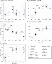
          Thirty-four wild-type mice (129S1/SvlmJ) were anesthetized and imaged between postnatal (P) day 7 and P21. OCT instrumentation was developed to optimize signal intensity and image quality. Semi-automatic segmentation tools were developed to quantify the retinal thickness of the nerve fiber layer (NFL), inner plexiform layer (IPL), inner nuclear layer (INL), and the outer retinal layers (ORL), in addition to the total retina. The retinal maturation was characterized by comparing layer thicknesses between consecutive time points.

          Results

          From P7 to P10, the IPL increased significantly, consistent with retinal synaptogenesis. From P10 to P12, the IPL and ORL also increased, which is coherent with synaptic connectivity and photoreceptor maturation. In contrast, during these periods, the INL decreased significantly, consistent with cellular densification and selective apoptotic “pruning” of the tissue during nuclear migration. Thereafter from P12 to P21, the INL continued to thin (significantly from P17 to P21) whereas the other layers remained unchanged. No time-dependent changes were observed in the NFL. Overall, changes in the total retina were attributed to those in the IPL, INL, and ORL. Regions of the retina adjacent to the optic nerve head were thinner than distal regions during maturation.

          Conclusions

          Changes in retinal layer thickness are consistent with retinal developmental mechanisms. Accordingly, this report opens new horizons in using our system in the mouse to characterize longitudinally developmental digressions in models of human diseases.

          Related collections

          Most cited references92

          • Record: found
          • Abstract: found
          • Article: not found

          NIH Image to ImageJ: 25 years of image analysis

          For the past twenty five years the NIH family of imaging software, NIH Image and ImageJ have been pioneers as open tools for scientific image analysis. We discuss the origins, challenges and solutions of these two programs, and how their history can serve to advise and inform other software projects.
            Bookmark
            • Record: found
            • Abstract: found
            • Article: not found

            Proposed lexicon for anatomic landmarks in normal posterior segment spectral-domain optical coherence tomography: the IN•OCT consensus.

            To develop a consensus nomenclature for the classification of retinal and choroidal layers and bands visible on spectral-domain optical coherence tomography (SD-OCT) images of a normal eye.
              Bookmark
              • Record: found
              • Abstract: found
              • Article: not found

              Anatomical correlates to the bands seen in the outer retina by optical coherence tomography: literature review and model.

              To evaluate the validity of commonly used anatomical designations for the four hyperreflective outer retinal bands seen in current-generation optical coherence tomography, a scale model of outer retinal morphology was created using published information for direct comparison with optical coherence tomography scans. Articles and books concerning histology of the outer retina from 1900 until 2009 were evaluated, and data were used to create a scale model drawing. Boundaries between outer retinal tissue compartments described by the model were compared with intensity variations of representative spectral-domain optical coherence tomography scans using longitudinal reflectance profiles to determine the region of origin of the hyperreflective outer retinal bands. This analysis showed a high likelihood that the spectral-domain optical coherence tomography bands attributed to the external limiting membrane (the first, innermost band) and to the retinal pigment epithelium (the fourth, outermost band) are correctly attributed. Comparative analysis showed that the second band, often attributed to the boundary between inner and outer segments of the photoreceptors, actually aligns with the ellipsoid portion of the inner segments. The third band corresponded to an ensheathment of the cone outer segments by apical processes of the retinal pigment epithelium in a structure known as the contact cylinder. Anatomical attributions and subsequent pathophysiologic assessments pertaining to the second and third outer retinal hyperreflective bands may not be correct. This analysis has identified testable hypotheses for the actual correlates of the second and third bands. Nonretinal pigment epithelium contributions to the fourth band (e.g., Bruch membrane) remain to be determined.
                Bookmark

                Author and article information

                Journal
                Invest Ophthalmol Vis Sci
                Invest Ophthalmol Vis Sci
                iovs
                IOVS
                Investigative Ophthalmology & Visual Science
                The Association for Research in Vision and Ophthalmology
                0146-0404
                1552-5783
                26 October 2021
                October 2021
                : 62
                : 13
                : 20
                Affiliations
                [1 ]Institute of Biomedical Engineering, University of Montréal, Montréal, Canada
                [2 ]Research Center, CHU Sainte-Justine, Montréal, Canada
                [3 ]Department of Pharmacology, University of Montréal, Montréal, Canada
                [4 ]Department of Pediatrics, University of Montréal, Montréal, Canada
                [5 ]Department of Ophthalmology, University of Montréal, Montréal, Canada
                [6 ]Department of Engineering Physics, Polytechnique Montréal, Montréal, Canada
                [7 ]Department of Radiology, Radio-oncology and Nuclear Medicine, University of Montréal, Montréal, Canada
                Author notes
                Correspondence: Mathieu Dehaes, 3175 Cote Ste-Catherine, Montréal, H3T 1C5, Canada; mathieu.dehaes@ 123456umontreal.ca .
                Article
                IOVS-19-28941
                10.1167/iovs.62.13.20
                8556565
                34698774
                7387fad8-919a-41b0-a484-4228ed4886fc
                Copyright 2021 The Authors

                This work is licensed under a Creative Commons Attribution 4.0 International License.

                History
                : 23 September 2021
                : 18 December 2019
                Page count
                Pages: 12
                Categories
                Retina
                Retina

                retina,development,microstructure,wild-type mouse,optical coherence tomography (oct)

                Comments

                Comment on this article