7
views
0
recommends
+1 Recommend
0 collections
    0
    shares
      • Record: found
      • Abstract: found
      • Article: found
      Is Open Access

      Estimation of Diabetic Retinal Microaneurysm Perfusion Parameters Based on Computational Fluid Dynamics Modeling of Adaptive Optics Scanning Laser Ophthalmoscopy

      research-article

      Read this article at

      Bookmark
          There is no author summary for this article yet. Authors can add summaries to their articles on ScienceOpen to make them more accessible to a non-specialist audience.

          Abstract

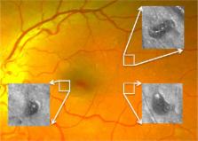
          Diabetic retinopathy (DR) is a leading cause of vision loss worldwide. Microaneurysms (MAs), which are abnormal outpouchings of the retinal vessels, are early and hallmark lesions of DR. The presence and severity of MAs are utilized to determine overall DR severity. In addition, MAs can directly contribute to retinal neural pathology by leaking fluid into the surrounding retina, causing abnormal central retinal thickening and thereby frequently leading to vision loss. Vascular perfusion parameters such as shear rate (SR) or wall shear stress (WSS) have been linked to blood clotting and endothelial cell dysfunction, respectively in non-retinal vasculature. However, despite the importance of MAs as a key aspect of diabetic retinal pathology, much remains unknown as to how structural characteristics of individual MAs are associated with these perfusion attributes. MA structural information obtained on high resolution adaptive optics scanning laser ophthalmoscopy (AOSLO) was utilized to estimate perfusion parameters through Computational Fluid Dynamics (CFD) analysis of the AOSLO images. The HemeLB flow solver was used to simulate steady-state and time-dependent fluid flow using both commodity hospital-based and high performance computing resources, depending on the degree of detail required in the simulations. Our results indicate that WSS is lowest in MA regions furthest away from the feeding vessels. Furthermore, areas of low SR are associated with clot location in saccular MAs. These findings suggest that morphology and CFD estimation of perfusion parameters may be useful tools for determining the likelihood of clot presence in individual diabetic MAs.

          Related collections

          Most cited references32

          • Record: found
          • Abstract: found
          • Article: not found

          Grading diabetic retinopathy from stereoscopic color fundus photographs--an extension of the modified Airlie House classification. ETDRS report number 10. Early Treatment Diabetic Retinopathy Study Research Group.

          (1991)
          The modified Airlie House classification of diabetic retinopathy has been extended for use in the Early Treatment Diabetic Retinopathy Study (ETDRS). The revised classification provides additional steps in the grading scale for some characteristics, separates other characteristics previously combined, expands the section on macular edema, and adds several characteristics not previously graded. The classification is described and illustrated and its reproducibility between graders is assessed by calculating percentages of agreement and kappa statistics for duplicate gradings of baseline color nonsimultaneous stereoscopic fundus photographs. For retinal hemorrhages and/or microaneurysms, hard exudates, new vessels, fibrous proliferations, and macular edema, agreement was substantial (weighted kappa, 0.61 to 0.80). For soft exudates, intraretinal microvascular abnormalities, and venous beading, agreement was moderate (weighted kappa, 0.41 to 0.60). A double grading system, with adjudication of disagreements of two or more steps between duplicate gradings, led to some improvement in reproducibility for most characteristics.
            Bookmark
            • Record: found
            • Abstract: not found
            • Article: not found

            Lattice-Boltzmann Method for Complex Flows

              Bookmark
              • Record: found
              • Abstract: found
              • Article: not found

              Fundus photographic risk factors for progression of diabetic retinopathy. ETDRS report number 12. Early Treatment Diabetic Retinopathy Study Research Group.

              (1991)
              In the Early Treatment Diabetic Retinopathy Study, a randomized clinical trial sponsored by the National Eye Institute, one eye of each patient was assigned to early photocoagulation and the other to deferral of photocoagulation (i.e., careful follow-up and initiation of photocoagulation only if high-risk proliferative retinopathy developed). This design allowed observation of the natural course of diabetic retinopathy in the initially untreated eye. Gradings of baseline stereoscopic fundus photographs of eyes with nonproliferative retinopathy assigned to deferral of photocoagulation were used to examine the power of various abnormalities and combinations of abnormalities to predict progression to proliferative retinopathy in photographs taken at the 1-, 3-, and 5-year follow-up visits. Severity of intraretinal microvascular abnormalities, hemorrhages and/or microaneurysms, and venous beading were found to be the most important factors in predicting progression. On the basis of these analyses and other considerations, a retinopathy severity scale was developed. This scale, which divides diabetic retinopathy into 13 levels ranging from absence of retinopathy to severe vitreous hemorrhage, can be used to describe overall retinopathy severity and change in severity over time.
                Bookmark

                Author and article information

                Contributors
                Journal
                Front Physiol
                Front Physiol
                Front. Physiol.
                Frontiers in Physiology
                Frontiers Media S.A.
                1664-042X
                07 September 2018
                2018
                : 9
                : 989
                Affiliations
                [1] 1Centre for Medical Informatics, Usher Institute, The University of Edinburgh , Edinburgh, United Kingdom
                [2] 2Beetham Eye Institute, Joslin Diabetes Center , Boston, MA, United States
                [3] 3Department of Ophthalmology, Harvard Medical School , Boston, MA, United States
                Author notes

                Edited by: Peter V. Coveney, University College London, United Kingdom

                Reviewed by: Paolo Melillo, Università degli Studi della Campania “Luigi Vanvitelli” Caserta, Italy; Mariano Vázquez, Barcelona Supercomputing Center, Spain; Jazmin Aguado-Sierra, Barcelona Supercomputing Center, Spain

                *Correspondence: Jennifer K. Sun jennifer.sun@ 123456joslin.harvard.edu

                This article was submitted to Computational Physiology and Medicine, a section of the journal Frontiers in Physiology

                †These authors have contributed equally to this work

                Article
                10.3389/fphys.2018.00989
                6137139
                73c86f78-d04c-4760-b68a-00f6d843573b
                Copyright © 2018 Bernabeu, Lu, Abu-Qamar, Aiello and Sun.

                This is an open-access article distributed under the terms of the Creative Commons Attribution License (CC BY). The use, distribution or reproduction in other forums is permitted, provided the original author(s) and the copyright owner(s) are credited and that the original publication in this journal is cited, in accordance with accepted academic practice. No use, distribution or reproduction is permitted which does not comply with these terms.

                History
                : 15 December 2017
                : 05 July 2018
                Page count
                Figures: 13, Tables: 1, Equations: 0, References: 45, Pages: 11, Words: 6695
                Funding
                Funded by: National Eye Institute 10.13039/100000053
                Award ID: 1R01EY0-24702-01
                Award ID: 2R44EY-16295-04A1
                Funded by: Juvenile Diabetes Research Foundation 10.13039/100008664
                Award ID: 2-SRA-2014-264-M-R
                Award ID: 17-2011-359
                Funded by: Engineering and Physical Sciences Research Council 10.13039/501100000266
                Award ID: EP/L00030X/1
                Funded by: Dubai Harvard Foundation for Medical Research 10.13039/100011886
                Funded by: Massachusetts Lions Eye Research Fund 10.13039/100007745
                Funded by: Fondation Leducq 10.13039/501100001674
                Award ID: 17 CVD 03
                Categories
                Physiology
                Original Research

                Anatomy & Physiology
                diabetic retinopathy,microaneurysm,adaptive optics,blood flow,computational fluid dynamics

                Comments

                Comment on this article