41
views
0
recommends
+1 Recommend
0 collections
    0
    shares
      • Record: found
      • Abstract: found
      • Article: found
      Is Open Access

      State-dependent block of Orai3 TM1 and TM3 cysteine mutants: Insights into 2-APB activation

      research-article

      Read this article at

      Bookmark
          There is no author summary for this article yet. Authors can add summaries to their articles on ScienceOpen to make them more accessible to a non-specialist audience.

          Abstract

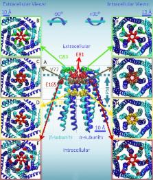
          Residue E165, in transmembrane helix 3, participates in formation of the dilated pore of the 2-APB–activated Orai3 channel but not that of the more selective store-operated Orai3 pore.

          Abstract

          After endoplasmic reticulum (ER) Ca 2+ store depletion, Orai channels in the plasma membrane (PM) are activated directly by ER-resident stromal interacting molecule (STIM) proteins to form the Ca 2+-selective Ca 2+ release-activated Ca 2+ (CRAC) channel. Of the three human Orai channel homologues, only Orai3 can be activated by high concentrations (>50 µM) of 2-aminoethyl diphenylborinate (2-APB). 2-APB activation of Orai3 occurs without STIM1–Orai3 interaction or store depletion, and results in a cationic, nonselective current characterized by biphasic inward and outward rectification. Here we use cysteine scanning mutagenesis, thiol-reactive reagents, and patch-clamp analysis to define the residues that assist in formation of the 2-APB–activated Orai3 pore. Mutating transmembrane (TM) 1 residues Q83, V77, and L70 to cysteine results in potentiated block by cadmium ions (Cd 2+). TM1 mutants E81C, G73A, G73C, and R66C form channels that are not sensitive to 2-APB activation. We also find that Orai3 mutant V77C is sensitive to block by 2-aminoethyl methanethiosulfonate (MTSEA), but not 2-(trimethylammonium)ethyl methanethiosulfonate (MTSET). Block induced by reaction with MTSEA is state dependent, as it occurs only when Orai3-V77C channels are opened by either 2-APB or by cotransfection with STIM1 and concurrent passive store depletion. We also analyzed TM3 residue E165. Mutation E165A in Orai3 results in diminished 2-APB–activated currents. However, it has little effect on store-operated current density. Furthermore, mutation E165C results in Cd 2+-induced block that is state dependent: Cd 2+ only blocks 2-APB–activated, not store-operated, mutant channels. Our data suggest that the dilated pore of 2-APB–activated Orai3 is lined by TM1 residues, but also allows for TM3 E165 to approach the central axis of the channel that forms the conducting pathway, or pore.

          Related collections

          Most cited references31

          • Record: found
          • Abstract: found
          • Article: not found

          A mutation in Orai1 causes immune deficiency by abrogating CRAC channel function.

          Antigen stimulation of immune cells triggers Ca2+ entry through Ca2+ release-activated Ca2+ (CRAC) channels, promoting the immune response to pathogens by activating the transcription factor NFAT. We have previously shown that cells from patients with one form of hereditary severe combined immune deficiency (SCID) syndrome are defective in store-operated Ca2+ entry and CRAC channel function. Here we identify the genetic defect in these patients, using a combination of two unbiased genome-wide approaches: a modified linkage analysis with single-nucleotide polymorphism arrays, and a Drosophila RNA interference screen designed to identify regulators of store-operated Ca2+ entry and NFAT nuclear import. Both approaches converged on a novel protein that we call Orai1, which contains four putative transmembrane segments. The SCID patients are homozygous for a single missense mutation in ORAI1, and expression of wild-type Orai1 in SCID T cells restores store-operated Ca2+ influx and the CRAC current (I(CRAC)). We propose that Orai1 is an essential component or regulator of the CRAC channel complex.
            Bookmark
            • Record: found
            • Abstract: found
            • Article: not found

            STIM is a Ca2+ sensor essential for Ca2+-store-depletion-triggered Ca2+ influx.

            Ca(2+) signaling in nonexcitable cells is typically initiated by receptor-triggered production of inositol-1,4,5-trisphosphate and the release of Ca(2+) from intracellular stores. An elusive signaling process senses the Ca(2+) store depletion and triggers the opening of plasma membrane Ca(2+) channels. The resulting sustained Ca(2+) signals are required for many physiological responses, such as T cell activation and differentiation. Here, we monitored receptor-triggered Ca(2+) signals in cells transfected with siRNAs against 2,304 human signaling proteins, and we identified two proteins required for Ca(2+)-store-depletion-mediated Ca(2+) influx, STIM1 and STIM2. These proteins have a single transmembrane region with a putative Ca(2+) binding domain in the lumen of the endoplasmic reticulum. Ca(2+) store depletion led to a rapid translocation of STIM1 into puncta that accumulated near the plasma membrane. Introducing a point mutation in the STIM1 Ca(2+) binding domain resulted in prelocalization of the protein in puncta, and this mutant failed to respond to store depletion. Our study suggests that STIM proteins function as Ca(2+) store sensors in the signaling pathway connecting Ca(2+) store depletion to Ca(2+) influx.
              Bookmark
              • Record: found
              • Abstract: found
              • Article: not found

              Store-operated calcium channels.

              In electrically nonexcitable cells, Ca(2+) influx is essential for regulating a host of kinetically distinct processes involving exocytosis, enzyme control, gene regulation, cell growth and proliferation, and apoptosis. The major Ca(2+) entry pathway in these cells is the store-operated one, in which the emptying of intracellular Ca(2+) stores activates Ca(2+) influx (store-operated Ca(2+) entry, or capacitative Ca(2+) entry). Several biophysically distinct store-operated currents have been reported, but the best characterized is the Ca(2+) release-activated Ca(2+) current, I(CRAC). Although it was initially considered to function only in nonexcitable cells, growing evidence now points towards a central role for I(CRAC)-like currents in excitable cells too. In spite of intense research, the signal that relays the store Ca(2+) content to CRAC channels in the plasma membrane, as well as the molecular identity of the Ca(2+) sensor within the stores, remains elusive. Resolution of these issues would be greatly helped by the identification of the CRAC channel gene. In some systems, evidence suggests that store-operated channels might be related to TRP homologs, although no consensus has yet been reached. Better understood are mechanisms that inactivate store-operated entry and hence control the overall duration of Ca(2+) entry. Recent work has revealed a central role for mitochondria in the regulation of I(CRAC), and this is particularly prominent under physiological conditions. I(CRAC) therefore represents a dynamic interplay between endoplasmic reticulum, mitochondria, and plasma membrane. In this review, we describe the key electrophysiological features of I(CRAC) and other store-operated Ca(2+) currents and how they are regulated, and we consider recent advances that have shed insight into the molecular mechanisms involved in this ubiquitous and vital Ca(2+) entry pathway.
                Bookmark

                Author and article information

                Journal
                J Gen Physiol
                J. Gen. Physiol
                jgp
                jgp
                The Journal of General Physiology
                The Rockefeller University Press
                0022-1295
                1540-7748
                May 2014
                : 143
                : 5
                : 621-631
                Affiliations
                Department of Physiology and Biophysics, University of California, Irvine, Irvine, CA 92697
                Author notes
                Correspondence to Michael D. Cahalan: mcahalan@ 123456uci.edu
                Article
                201411171
                10.1085/jgp.201411171
                4003185
                24733836
                770729b2-5668-48c5-a83c-b0aa5de063a2
                © 2014 Amcheslavsky et al.

                This article is distributed under the terms of an Attribution–Noncommercial–Share Alike–No Mirror Sites license for the first six months after the publication date (see http://www.rupress.org/terms). After six months it is available under a Creative Commons License (Attribution–Noncommercial–Share Alike 3.0 Unported license, as described at http://creativecommons.org/licenses/by-nc-sa/3.0/).

                History
                : 23 January 2014
                : 24 March 2014
                Categories
                Research Articles

                Anatomy & Physiology
                Anatomy & Physiology

                Comments

                Comment on this article