33
views
0
recommends
+1 Recommend
0 collections
    0
    shares
      • Record: found
      • Abstract: found
      • Article: found
      Is Open Access

      Regulation of ENaC-Mediated Sodium Reabsorption by Peroxisome Proliferator-Activated Receptors

      review-article

      Read this article at

      Bookmark
          There is no author summary for this article yet. Authors can add summaries to their articles on ScienceOpen to make them more accessible to a non-specialist audience.

          Abstract

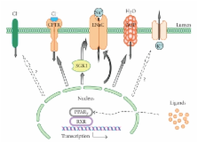
          Peroxisome proliferator-activated receptors (PPARs) are members of a steroid hormone receptor superfamily that responds to changes in lipid and glucose homeostasis. Peroxisomal proliferator-activated receptor subtype γ (PPAR γ) has received much attention as the target for antidiabetic drugs, as well as its role in responding to endogenous compounds such as prostaglandin J 2. However, thiazolidinediones (TZDs), the synthetic agonists of the PPAR γ are tightly associated with fluid retention and edema, as potentially serious side effects. The epithelial sodium channel (ENaC) represents the rate limiting step for sodium absorption in the renal collecting duct. Consequently, ENaC is a central effector impacting systemic blood volume and pressure. The role of PPAR γ agonists on ENaC activity remains controversial. While PPAR γ agonists were shown to stimulate ENaC-mediated renal salt absorption, probably via Serum- and Glucocorticoid-Regulated Kinase 1 (SGK1), other studies reported that PPAR γ agonist-induced fluid retention is independent of ENaC activity. The current paper provides new insights into the control and function of ENaC and ENaC-mediated sodium transport as well as several other epithelial channels/transporters by PPARs and particularly PPAR γ. The potential contribution of arachidonic acid (AA) metabolites in PPAR-dependent mechanisms is also discussed.

          Related collections

          Most cited references89

          • Record: found
          • Abstract: not found
          • Article: not found

          Thiazolidinediones

            Bookmark
            • Record: found
            • Abstract: found
            • Article: not found

            Differential expression of peroxisome proliferator-activated receptors (PPARs): tissue distribution of PPAR-alpha, -beta, and -gamma in the adult rat.

            Peroxisome proliferator-activated receptors (PPARs) are members of the nuclear hormone receptor superfamily that can be activated by various xenobiotics and natural fatty acids. These transcription factors primarily regulate genes involved in lipid metabolism and also play a role in adipocyte differentiation. We present the expression patterns of the PPAR subtypes in the adult rat, determined by in situ hybridization using specific probes for PPAR-alpha, -beta and -gamma, and by immunohistochemistry using a polyclonal antibody that recognizes the three rat PPAR subtypes. In numerous cell types from either ectodermal, mesodermal, or endodermal origin, PPARs are coexpressed, with relative levels varying between them from one cell type to the other. PPAR-alpha is highly expressed in hepatocytes, cardiomyocytes, enterocytes, and the proximal tubule cells of kidney. PPAR-beta is expressed ubiquitously and often at higher levels than PPAR-alpha and -gamma. PPAR-gamma is expressed predominantly in adipose tissue and the immune system. Our results suggest new potential directions to investigate the functions of the different PPAR subtypes.
              Bookmark
              • Record: found
              • Abstract: found
              • Article: not found

              The organization, promoter analysis, and expression of the human PPARgamma gene.

              PPARgamma is a member of the PPAR subfamily of nuclear receptors. In this work, the structure of the human PPARgamma cDNA and gene was determined, and its promoters and tissue-specific expression were functionally characterized. Similar to the mouse, two PPAR isoforms, PPARgamma1 and PPARgamma2, were detected in man. The relative expression of human PPARgamma was studied by a newly developed and sensitive reverse transcriptase-competitive polymerase chain reaction method, which allowed us to distinguish between PPARgamma1 and gamma2 mRNA. In all tissues analyzed, PPARgamma2 was much less abundant than PPARgamma1. Adipose tissue and large intestine have the highest levels of PPARgamma mRNA; kidney, liver, and small intestine have intermediate levels; whereas PPARgamma is barely detectable in muscle. This high level expression of PPARgamma in colon warrants further study in view of the well established role of fatty acid and arachidonic acid derivatives in colonic disease. Similarly as mouse PPARgammas, the human PPARgammas are activated by thiazolidinediones and prostaglandin J and bind with high affinity to a PPRE. The human PPARgamma gene has nine exons and extends over more than 100 kilobases of genomic DNA. Alternate transcription start sites and alternate splicing generate the PPARgamma1 and PPARgamma2 mRNAs, which differ at their 5'-ends. PPARgamma1 is encoded by eight exons, and PPARgamma2 is encoded by seven exons. The 5'-untranslated sequence of PPARgamma1 is comprised of exons A1 and A2, whereas that of PPARgamma2 plus the additional PPARgamma2-specific N-terminal amino acids are encoded by exon B, located between exons A2 and A1. The remaining six exons, termed 1 to 6, are common to the PPARgamma1 and gamma2. Knowledge of the gene structure will allow screening for PPARgamma mutations in humans with metabolic disorders, whereas knowledge of its expression pattern and factors regulating its expression could be of major importance in understanding its biology.
                Bookmark

                Author and article information

                Journal
                PPAR Res
                PPAR
                PPAR Research
                Hindawi Publishing Corporation
                1687-4757
                1687-4765
                2010
                14 June 2010
                : 2010
                : 703735
                Affiliations
                1Department of Physiology, Medical College of Wisconsin, Milwaukee, WI 53226, USA
                2Department of Pharmacology and Toxicology, Medical College of Wisconsin, Milwaukee, WI 53226, USA
                3Cardiovascular Research Center, Medical College of Wisconsin, Milwaukee, WI 53226, USA
                4Kidney Disease Center, Medical College of Wisconsin, Milwaukee, WI 53226, USA
                Author notes
                *Alexander Staruschenko: staruschenko@ 123456mcw.edu

                Academic Editor: Tianxin Yang

                Article
                10.1155/2010/703735
                2896859
                20613963
                7e50195b-f9f3-41f6-b376-28df0d4cc3f8
                Copyright © 2010 Tengis S. Pavlov et al.

                This is an open access article distributed under the Creative Commons Attribution License, which permits unrestricted use, distribution, and reproduction in any medium, provided the original work is properly cited.

                History
                : 6 January 2010
                : 16 March 2010
                : 14 April 2010
                Categories
                Review Article

                Biochemistry
                Biochemistry

                Comments

                Comment on this article