15
views
0
recommends
+1 Recommend
0 collections
    0
    shares
      • Record: found
      • Abstract: found
      • Article: found
      Is Open Access

      Globular Adiponectin Causes Tolerance to LPS-Induced TNF-α Expression via Autophagy Induction in RAW 264.7 Macrophages: Involvement of SIRT1/FoxO3A Axis

      research-article
      , , , *
      PLoS ONE
      Public Library of Science

      Read this article at

      Bookmark
          There is no author summary for this article yet. Authors can add summaries to their articles on ScienceOpen to make them more accessible to a non-specialist audience.

          Abstract

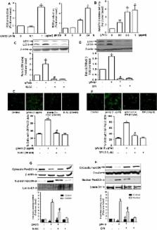
          Adiponectin, an adipokine predominantly produced from adipose tissue, exhibited potent anti-inflammatory properties. In particular, it inhibits production of pro-inflammatory cytokines, including tumor necrosis factor-α (TNF-α), in macrophages. Autophagy, an intracellular self-digestion process, has been recently shown to regulate inflammatory responses. In the present study, we investigated the role of autophagy induction in the suppression of Lipopolysaccharide (LPS) -induced TNF-α expression by globular adiponectin (gAcrp) and its potential mechanisms. Herein, we found that gAcrp treatment increased expression of genes related with autophagy, including Atg5 and microtubule-associated protein light chain (LC3B), induced autophagosome formation and autophagy flux in RAW 264.7 macrophages. Similar results were observed in primary macrophages isolated peritoneum of mice. Interestingly, inhibition of autophagy by pretreatment with Bafilomycin A1 or knocking down of LC3B gene restored suppression of TNF-α expression, tumor necrosis factor receptor- associated factor 6 (TRAF6) expression and p38MAPK phosphorylation by gAcrp, implying a critical role of autophagy induction in the development of tolerance to LPS-induced TNF-α expression by gAcrp. We also found that knocking-down of FoxO3A, a forkhead box O member of transcription factor, blocked gAcrp-induced expression of LC3II and Atg5. Moreover, gene silencing of Silent information regulator 1 (SIRT1) blocked both gAcrp-induced nuclear translocation of FoxO3A and LC3II expression. Finally, pretreatment with ROS inhibitors, prevented gAcrp-induced SIRT1 expression and further generated inhibitory effects on gAcrp-induced autophagy, indicating a role of ROS production in gAcrp-induced SIRT1 expression and subsequent autophagy induction. Taken together, these findings indicate that globular adiponectin suppresses LPS-induced TNF-α expression, at least in part, via autophagy activation. Furthermore, SIRT1-FoxO3A axis plays a crucial role in gAcrp-induced autophagy in macrophages.

          Related collections

          Most cited references53

          • Record: found
          • Abstract: found
          • Article: not found
          Is Open Access

          Decoding cell death signals in liver inflammation.

          Inflammation can be either beneficial or detrimental to the liver, depending on multiple factors. Mild (i.e., limited in intensity and destined to resolve) inflammatory responses have indeed been shown to exert consistent hepatoprotective effects, contributing to tissue repair and promoting the re-establishment of homeostasis. Conversely, excessive (i.e., disproportionate in intensity and permanent) inflammation may induce a massive loss of hepatocytes and hence exacerbate the severity of various hepatic conditions, including ischemia-reperfusion injury, systemic metabolic alterations (e.g., obesity, diabetes, non-alcoholic fatty liver disorders), alcoholic hepatitis, intoxication by xenobiotics and infection, de facto being associated with irreversible liver damage, fibrosis, and carcinogenesis. Both liver-resident cells (e.g., Kupffer cells, hepatic stellate cells, sinusoidal endothelial cells) and cells that are recruited in response to injury (e.g., monocytes, macrophages, dendritic cells, natural killer cells) emit pro-inflammatory signals including - but not limited to - cytokines, chemokines, lipid messengers, and reactive oxygen species that contribute to the apoptotic or necrotic demise of hepatocytes. In turn, dying hepatocytes release damage-associated molecular patterns that-upon binding to evolutionary conserved pattern recognition receptors-activate cells of the innate immune system to further stimulate inflammatory responses, hence establishing a highly hepatotoxic feedforward cycle of inflammation and cell death. In this review, we discuss the cellular and molecular mechanisms that account for the most deleterious effect of hepatic inflammation at the cellular level, that is, the initiation of a massive cell death response among hepatocytes. Copyright © 2013 European Association for the Study of the Liver. Published by Elsevier B.V. All rights reserved.
            Bookmark
            • Record: found
            • Abstract: found
            • Article: found
            Is Open Access

            Effects of obesity on bone metabolism

            Jay Cao (2011)
            Obesity is traditionally viewed to be beneficial to bone health because of well-established positive effect of mechanical loading conferred by body weight on bone formation, despite being a risk factor for many other chronic health disorders. Although body mass has a positive effect on bone formation, whether the mass derived from an obesity condition or excessive fat accumulation is beneficial to bone remains controversial. The underline pathophysiological relationship between obesity and bone is complex and continues to be an active research area. Recent data from epidemiological and animal studies strongly support that fat accumulation is detrimental to bone mass. To our knowledge, obesity possibly affects bone metabolism through several mechanisms. Because both adipocytes and osteoblasts are derived from a common multipotential mesenchymal stem cell, obesity may increase adipocyte differentiation and fat accumulation while decrease osteoblast differentiation and bone formation. Obesity is associated with chronic inflammation. The increased circulating and tissue proinflammatory cytokines in obesity may promote osteoclast activity and bone resorption through modifying the receptor activator of NF-κB (RANK)/RANK ligand/osteoprotegerin pathway. Furthermore, the excessive secretion of leptin and/or decreased production of adiponectin by adipocytes in obesity may either directly affect bone formation or indirectly affect bone resorption through up-regulated proinflammatory cytokine production. Finally, high-fat intake may interfere with intestinal calcium absorption and therefore decrease calcium availability for bone formation. Unraveling the relationship between fat and bone metabolism at molecular level may help us to develop therapeutic agents to prevent or treat both obesity and osteoporosis. Obesity, defined as having a body mass index ≥ 30 kg/m2, is a condition in which excessive body fat accumulates to a degree that adversely affects health [1]. The rates of obesity rates have doubled since 1980 [2] and as of 2007, 33% of men and 35% of women in the US are obese [3]. Obesity is positively associated to many chronic disorders such as hypertension, dyslipidemia, type 2 diabetes mellitus, coronary heart disease, and certain cancers [4-6]. It is estimated that the direct medical cost associated with obesity in the United States is ~$100 billion per year [7]. Bone mass and strength decrease during adulthood, especially in women after menopause [8]. These changes can culminate in osteoporosis, a disease characterized by low bone mass and microarchitectural deterioration resulting in increased bone fracture risk. It is estimated that there are about 10 million Americans over the age of 50 who have osteoporosis while another 34 million people are at risk of developing the disease [9]. In 2001, osteoporosis alone accounted for some $17 billion in direct annual healthcare expenditure. Several lines of evidence suggest that obesity and bone metabolism are interrelated. First, both osteoblasts (bone forming cells) and adipocytes (energy storing cells) are derived from a common mesenchymal stem cell [10] and agents inhibiting adipogenesis stimulated osteoblast differentiation [11-13] and vice versa, those inhibiting osteoblastogenesis increased adipogenesis [14]. Second, decreased bone marrow osteoblastogenesis with aging is usually accompanied with increased marrow adipogenesis [15,16]. Third, chronic use of steroid hormone, such as glucocorticoid, results in obesity accompanied by rapid bone loss [17,18]. Fourth, both obesity and osteoporosis are associated with elevated oxidative stress and increased production of proinflammatory cytokines [19,20]. At present, the mechanisms for the effects of obesity on bone metabolism are not well defined and will be the focus of this review.
              Bookmark
              • Record: found
              • Abstract: found
              • Article: not found

              Adiponectin, a new member of the family of soluble defense collagens, negatively regulates the growth of myelomonocytic progenitors and the functions of macrophages.

              We investigated the functions of adiponectin, an adipocyte-specific secretory protein and a new member of the family of soluble defense collagens, in hematopoiesis and immune responses. Adiponectin suppressed colony formation from colony-forming units (CFU)-granulocyte-macrophage, CFU-macrophage, and CFU-granulocyte, whereas it had no effect on that of burst-forming units-erythroid or mixed erythroid-myeloid CFU. In addition, adiponectin inhibited proliferation of 4 of 9 myeloid cell lines but did not suppress proliferation of erythroid or lymphoid cell lines except for one cell line. These results suggest that adiponectin predominantly inhibits proliferation of myelomonocytic lineage cells. At least one mechanism of the growth inhibition is induction of apoptosis because treatment of acute myelomonocytic leukemia lines with adiponectin induced the appearance of subdiploid peaks and oligonucleosomal DNA fragmentation. Aside from inhibiting growth of myelomonocytic progenitors, adiponectin suppressed mature macrophage functions. Treatment of cultured macrophages with adiponectin significantly inhibited their phagocytic activity and their lipopolysaccharide-induced production of tumor necrosis factor alpha. Suppression of phagocytosis by adiponectin is mediated by one of the complement C1q receptors, C1qRp, because this function was completely abrogated by the addition of an anti-C1qRp monoclonal antibody. These observations suggest that adiponectin is an important negative regulator in hematopoiesis and immune systems and raise the possibility that it may be involved in ending inflammatory responses through its inhibitory functions. (Blood. 2000;96:1723-1732)
                Bookmark

                Author and article information

                Contributors
                Role: Academic Editor
                Journal
                PLoS One
                PLoS ONE
                plos
                plosone
                PLoS ONE
                Public Library of Science (San Francisco, CA USA )
                1932-6203
                11 May 2015
                2015
                : 10
                : 5
                : e0124636
                Affiliations
                [001]College of Pharmacy, Yeungnam University, Gyeongsan, Gyeongsangbuk-do 712–749, Republic of Korea
                Northwestern University Feinberg School of Medicine, UNITED STATES
                Author notes

                Competing Interests: The authors have declared that no competing interests exist.

                Conceived and designed the experiments: NTP AS MJK PHP. Performed the experiments: NTP AS MJK. Analyzed the data: NTP AS PHP. Contributed reagents/materials/analysis tools: NTP PHP. Wrote the paper: NTP AS PHP.

                Article
                PONE-D-14-57166
                10.1371/journal.pone.0124636
                4427353
                25961287
                83cf4efd-69b7-4f4c-986d-b16e9e28c34d
                Copyright @ 2015

                This is an open access article distributed under the terms of the Creative Commons Attribution License, which permits unrestricted use, distribution, and reproduction in any medium, provided the original author and source are credited

                History
                : 20 December 2014
                : 17 March 2015
                Page count
                Figures: 8, Tables: 2, Pages: 22
                Funding
                This research was supported by the Basic Science Research Program through the National Research Foundation of Korea (NRF) funded by the Ministry of Education (2013R1A1A4A01011110). The funders had no role in study design, data collection and analysis, decision to publish, or preparation of the manuscript.
                Categories
                Research Article
                Custom metadata
                All relevant data are within the paper.

                Uncategorized
                Uncategorized

                Comments

                Comment on this article