2
views
0
recommends
+1 Recommend
0 collections
    0
    shares
      • Record: found
      • Abstract: found
      • Article: found
      Is Open Access

      Excessive Accumulation of Ca 2 + in Mitochondria of Y522S-RYR1 Knock-in Mice: A Link Between Leak From the Sarcoplasmic Reticulum and Altered Redox State

      research-article

      Read this article at

      Bookmark
          There is no author summary for this article yet. Authors can add summaries to their articles on ScienceOpen to make them more accessible to a non-specialist audience.

          Abstract

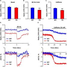
          Mice (Y522S or YS), carrying a mutation of the sarcoplasmic reticulum (SR) Ca 2+ release channel of skeletal muscle fibers (ryanodine receptor type-1, RyR1) which causes Ca 2+ leak, are a widely accepted and intensively studied model for human malignant hyperthermia (MH) susceptibility. Since the involvement of reactive oxygen species (ROS) and of mitochondria in MH crisis has been previously debated, here we sought to determine Ca 2+ uptake in mitochondria and its possible link with ROS production in single fibers isolated from flexor digitorum brevis (FDB) of YS mice. We found that Ca 2+ concentration in the mitochondrial matrix, as detected with the ratiometric FRET-based 4mtD3cpv probe, was higher in YS than in wild-type (WT) fibers at rest and after Ca 2+ release from SR during repetitive electrical stimulation or caffeine administration. Also mitochondrial ROS production associated with contractile activity (detected with Mitosox probe) was much higher in YS fibers than in WT. Importantly, the inhibition of mitochondrial Ca 2+ uptake achieved by silencing MCU reduced ROS accumulation in the matrix and Ca 2+ release from SR. Finally, inhibition of mitochondrial ROS accumulation using Mitotempo reduced SR Ca 2+ release in YS fibers exposed to caffeine. The present results support the view that mitochondria take up larger amounts of Ca 2+ in YS than in WT fibers and that mitochondrial ROS production substantially contributes to the increased caffeine-sensitivity and to the enhanced Ca 2+ release from SR in YS fibers.

          Related collections

          Most cited references40

          • Record: found
          • Abstract: found
          • Article: not found

          Mitochondria as sensors and regulators of calcium signalling.

          During the past two decades calcium (Ca(2+)) accumulation in energized mitochondria has emerged as a biological process of utmost physiological relevance. Mitochondrial Ca(2+) uptake was shown to control intracellular Ca(2+) signalling, cell metabolism, cell survival and other cell-type specific functions by buffering cytosolic Ca(2+) levels and regulating mitochondrial effectors. Recently, the identity of mitochondrial Ca(2+) transporters has been revealed, opening new perspectives for investigation and molecular intervention.
            Bookmark
            • Record: found
            • Abstract: found
            • Article: not found

            Regulation of mitochondrial dehydrogenases by calcium ions.

            Studies in Bristol in the 1960s and 1970s, led to the recognition that four mitochondrial dehydrogenases are activated by calcium ions. These are FAD-glycerol phosphate dehydrogenase, pyruvate dehydrogenase, NAD-isocitrate dehydrogenase and oxoglutarate dehydrogenase. FAD-glycerol phosphate dehydrogenase is located on the outer surface of the inner mitochondrial membrane and is influenced by changes in cytoplasmic calcium ion concentration. The other three enzymes are located within mitochondria and are regulated by changes in mitochondrial matrix calcium ion concentration. These and subsequent studies on purified enzymes, mitochondria and intact cell preparations have led to the widely accepted view that the activation of these enzymes is important in the stimulation of the respiratory chain and hence ATP supply under conditions of increased ATP demand in many stimulated mammalian cells. The effects of calcium ions on FAD-isocitrate dehydrogenase involve binding to an EF-hand binding motif within this enzyme but the binding sites involved in the effects of calcium ions on the three intramitochondrial dehydrogenases remain to be fully established. It is also emphasised in this article that these three dehydrogenases appear only to be regulated by calcium ions in vertebrates and that this raises some interesting and potentially important developmental issues.
              Bookmark
              • Record: found
              • Abstract: found
              • Article: not found

              Calcium at the Center of Cell Signaling: Interplay between Endoplasmic Reticulum, Mitochondria, and Lysosomes.

              In recent years, rapid discoveries have been made relating to Ca(2+) handling at specific organelles that have important implications for whole-cell Ca(2+) homeostasis. In particular, the structures of the endoplasmic reticulum (ER) Ca(2+) channels revealed by electron cryomicroscopy (cryo-EM), continuous updates on the structure, regulation, and role of the mitochondrial calcium uniporter (MCU) complex, and the analysis of lysosomal Ca(2+) signaling are milestones on the route towards a deeper comprehension of the complexity of global Ca(2+) signaling. In this review we summarize recent discoveries on the regulation of interorganellar Ca(2+) homeostasis and its role in pathophysiology.
                Bookmark

                Author and article information

                Contributors
                Journal
                Front Physiol
                Front Physiol
                Front. Physiol.
                Frontiers in Physiology
                Frontiers Media S.A.
                1664-042X
                13 September 2019
                2019
                : 10
                : 1142
                Affiliations
                [1] 1Department of Biomedical Sciences, School of Medicine and Surgery, University of Padova , Padua, Italy
                [2] 2Department of Biology, University of Padova , Padua, Italy
                [3] 3Center for Advanced Studies and Technology, Università degli Studi “G. d’Annunzio” Chieti–Pescara , Chieti, Italy
                [4] 4Department of Medicine and Aging Sciences, Università degli Studi “G. d’Annunzio” Chieti–Pescara , Chieti, Italy
                [5] 5Institute for Kinesiology Research, Science and Research Center of Koper , Koper, Slovenia
                Author notes

                Edited by: Susan V. Brooks, University of Michigan, United States

                Reviewed by: George G. Rodney, Baylor College of Medicine, United States; Paul D. Allen, University of Leeds, United Kingdom

                *Correspondence: Antonio Michelucci, antonio.michelucci@ 123456unich.it

                This article was submitted to Striated Muscle Physiology, a section of the journal Frontiers in Physiology

                Article
                10.3389/fphys.2019.01142
                6755340
                31607937
                83f9feac-374a-438e-ab77-44658369f6f5
                Copyright © 2019 Canato, Capitanio, Cancellara, Leanza, Raffaello, Vecellio Reane, Marcucci, Michelucci, Protasi and Reggiani.

                This is an open-access article distributed under the terms of the Creative Commons Attribution License (CC BY). The use, distribution or reproduction in other forums is permitted, provided the original author(s) and the copyright owner(s) are credited and that the original publication in this journal is cited, in accordance with accepted academic practice. No use, distribution or reproduction is permitted which does not comply with these terms.

                History
                : 31 December 2018
                : 21 August 2019
                Page count
                Figures: 8, Tables: 0, Equations: 0, References: 49, Pages: 13, Words: 0
                Categories
                Physiology
                Original Research

                Anatomy & Physiology
                excitation–contraction coupling,mitochondria,reactive oxygen species,ryanodine receptor,malignant hyperthermia

                Comments

                Comment on this article