Inviting an author to review:
Find an author and click ‘Invite to review selected article’ near their name.
Search for authorsSearch for similar articles
17
views
0
recommends
+1 Recommend
0 collections
    0
    shares
      • Record: found
      • Abstract: found
      • Article: found
      Is Open Access

      Contractile Properties of Esophageal Striated Muscle: Comparison with Cardiac and Skeletal Muscles in Rats

      research-article

      Read this article at

      Bookmark
          There is no author summary for this article yet. Authors can add summaries to their articles on ScienceOpen to make them more accessible to a non-specialist audience.

          Abstract

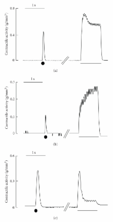
          The external muscle layer of the mammalian esophagus consists of striated muscles. We investigated the contractile properties of esophageal striated muscle by comparison with those of skeletal and cardiac muscles. Electrical field stimulation with single pulses evoked twitch-like contractile responses in esophageal muscle, similar to those in skeletal muscle in duration and similar to those in cardiac muscle in amplitude. The contractions of esophageal muscle were not affected by an inhibitor of gap junctions. Contractile responses induced by high potassium or caffeine in esophageal muscle were analogous to those in skeletal muscle. High-frequency stimulation induced a transient summation of contractions followed by sustained contractions with amplitudes similar to those of twitch-like contractions, although a large summation was observed in skeletal muscle. The results demonstrate that esophageal muscle has properties similar but not identical to those of skeletal muscle and that some specific properties may be beneficial for esophageal peristalsis.

          Related collections

          Most cited references43

          • Record: found
          • Abstract: found
          • Article: not found

          Structural and functional diversity of connexin genes in the mouse and human genome.

          Gap junctions are clustered channels between contacting cells through which direct intercellular communication via diffusion of ions and metabolites can occur. Two hemichannels, each built up of six connexin protein subunits in the plasma membrane of adjacent cells, can dock to each other to form conduits between cells. We have recently screened mouse and human genomic data bases and have found 19 connexin (Cx) genes in the mouse genome and 20 connexin genes in the human genome. One mouse connexin gene and two human connexin genes do not appear to have orthologs in the other genome. With three exceptions, the characterized connexin genes comprise two exons whereby the complete reading frame is located on the second exon. Targeted ablation of eleven mouse connexin genes revealed basic insights into the functional diversity of the connexin gene family. In addition, the phenotypes of human genetic disorders caused by mutated connexin genes further complement our understanding of connexin functions in the human organism. In this review we compare currently identified connexin genes in both the mouse and human genome and discuss the functions of gap junctions deduced from targeted mouse mutants and human genetic disorders.
            Bookmark
            • Record: found
            • Abstract: found
            • Article: not found

            Gap junctions: basic structure and function.

            Gap junctions allow the exchange of ions, second messengers, and small metabolites between adjacent cells and are formed by two unrelated protein families, the pannexins and connexins. Mutations in connexin genes cause a variety of genetic disorders, implicating a critical role in tissue homeostasis. Association of congenital skin disorders to mutations in different connexins has underscored the importance of gap junctional communication in the skin and its appendages. Here, we discuss the basic structure of gap junction channels and the function of connexin genes that have been associated with human disorders to explore the physiology of intercellular communication in skin.
              Bookmark
              • Record: found
              • Abstract: found
              • Article: not found

              Smooth muscle cell calcium activation mechanisms.

              Smooth muscle cell (SMC) contraction is controlled by the Ca2+ and Rho kinase signalling pathways. While the SMC Rho kinase system seems to be reasonably constant, there is enormous variation with regard to the mechanisms responsible for generating Ca2+ signals. One way of dealing with this diversity is to consider how this system has been adapted to control different SMC functions. Phasic SMCs (vas deferens, uterus and bladder) rely on membrane depolarization to drive Ca2+ influx across the plasma membrane. This depolarization can be induced by neurotransmitters or through the operation of a membrane oscillator. Many tonic SMCs (vascular, airway and corpus cavernosum) are driven by a cytosolic Ca2+ oscillator that generates periodic pulses of Ca2+. A similar oscillator is present in pacemaker cells such as the interstitial cells of Cajal (ICCs) and atypical SMCs that control other tonic SMCs (gastrointestinal, urethra, ureter). The changes in membrane potential induced by these cytosolic oscillators does not drive contraction directly but it functions to couple together individual oscillators to provide the synchronization that is a characteristic feature of many tonic SMCs.
                Bookmark

                Author and article information

                Journal
                J Biomed Biotechnol
                JBB
                Journal of Biomedicine and Biotechnology
                Hindawi Publishing Corporation
                1110-7243
                1110-7251
                2010
                1 April 2010
                : 2010
                : 459789
                Affiliations
                1Laboratory of Physiology, Department of Basic Veterinary Science, The United Graduate School of Veterinary Sciences, Gifu University, 1-1 Yanagido, Gifu 501-1193, Japan
                2Department of Applied Biology, Kyoto Institute of Technology, Matsugasaki, Sakyo, Kyoto 606-8585, Japan
                Author notes

                Academic Editor: Henk L. M. Granzier

                Article
                10.1155/2010/459789
                2850148
                20379364
                841d9398-e36d-44b1-8fc7-92c244202184
                Copyright © 2010 Takahiko Shiina et al.

                This is an open access article distributed under the Creative Commons Attribution License, which permits unrestricted use, distribution, and reproduction in any medium, provided the original work is properly cited.

                History
                : 30 October 2009
                : 1 February 2010
                Categories
                Research Article

                Molecular medicine
                Molecular medicine

                Comments

                Comment on this article