18
views
0
recommends
+1 Recommend
2 collections
    0
    shares
      • Record: found
      • Abstract: found
      • Article: found
      Is Open Access

      Secondary tension pneumothorax in a COVID-19 pneumonia patient: a case report

      research-article

      Read this article at

      Bookmark
          There is no author summary for this article yet. Authors can add summaries to their articles on ScienceOpen to make them more accessible to a non-specialist audience.

          Abstract

          Purpose

          Especially in elderly and multimorbid patients, Coronavirus Disease 2019 (COVID-19) may result in severe pneumonia and secondary complications. Recent studies showed pneumothorax in rare cases, but tension pneumothorax has only been reported once.

          Case presentation

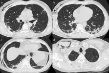
          A 47-year-old male was admitted to the emergency department with fever, dry cough and sore throat for the last 14 days as well as acute stenocardia and shortage of breath. Sputum testing (polymerase chain reaction, PCR) confirmed SARS-CoV-2 infection. Initial computed tomography (CT) showed bipulmonary groundglass opacities and consolidations with peripheral distribution. Hospitalization with supportive therapy (azithromycin) as well as non-invasive oxygenation led to a stabilization of the patient. After 5 days, sputum testing was negative and IgA/IgG antibody titres were positive for SARS-CoV-2. The patient was discharged after 7 days.

          On the 11th day, the patient realized pronounced dyspnoea after coughing and presented to the emergency department again. CT showed a right-sided tension pneumothorax, which was relieved by a chest drain (Buelau) via mini open thoracotomy. Negative pressure therapy resulted in regression of the pneumothorax and the patient was discharged after 9 days of treatment.

          Conclusion

          Treating physicians should be aware that COVID-19 patients might develop severe secondary pulmonary complications such as acute tension pneumothorax.

          Level of evidence

          V

          Electronic supplementary material

          The online version of this article (10.1007/s15010-020-01457-w) contains supplementary material, which is available to authorized users.

          Related collections

          Most cited references13

          • Record: found
          • Abstract: found
          • Article: not found

          A Novel Coronavirus from Patients with Pneumonia in China, 2019

          Summary In December 2019, a cluster of patients with pneumonia of unknown cause was linked to a seafood wholesale market in Wuhan, China. A previously unknown betacoronavirus was discovered through the use of unbiased sequencing in samples from patients with pneumonia. Human airway epithelial cells were used to isolate a novel coronavirus, named 2019-nCoV, which formed a clade within the subgenus sarbecovirus, Orthocoronavirinae subfamily. Different from both MERS-CoV and SARS-CoV, 2019-nCoV is the seventh member of the family of coronaviruses that infect humans. Enhanced surveillance and further investigation are ongoing. (Funded by the National Key Research and Development Program of China and the National Major Project for Control and Prevention of Infectious Disease in China.)
            Bookmark
            • Record: found
            • Abstract: found
            • Article: not found

            Epidemiological and clinical characteristics of 99 cases of 2019 novel coronavirus pneumonia in Wuhan, China: a descriptive study

            Summary Background In December, 2019, a pneumonia associated with the 2019 novel coronavirus (2019-nCoV) emerged in Wuhan, China. We aimed to further clarify the epidemiological and clinical characteristics of 2019-nCoV pneumonia. Methods In this retrospective, single-centre study, we included all confirmed cases of 2019-nCoV in Wuhan Jinyintan Hospital from Jan 1 to Jan 20, 2020. Cases were confirmed by real-time RT-PCR and were analysed for epidemiological, demographic, clinical, and radiological features and laboratory data. Outcomes were followed up until Jan 25, 2020. Findings Of the 99 patients with 2019-nCoV pneumonia, 49 (49%) had a history of exposure to the Huanan seafood market. The average age of the patients was 55·5 years (SD 13·1), including 67 men and 32 women. 2019-nCoV was detected in all patients by real-time RT-PCR. 50 (51%) patients had chronic diseases. Patients had clinical manifestations of fever (82 [83%] patients), cough (81 [82%] patients), shortness of breath (31 [31%] patients), muscle ache (11 [11%] patients), confusion (nine [9%] patients), headache (eight [8%] patients), sore throat (five [5%] patients), rhinorrhoea (four [4%] patients), chest pain (two [2%] patients), diarrhoea (two [2%] patients), and nausea and vomiting (one [1%] patient). According to imaging examination, 74 (75%) patients showed bilateral pneumonia, 14 (14%) patients showed multiple mottling and ground-glass opacity, and one (1%) patient had pneumothorax. 17 (17%) patients developed acute respiratory distress syndrome and, among them, 11 (11%) patients worsened in a short period of time and died of multiple organ failure. Interpretation The 2019-nCoV infection was of clustering onset, is more likely to affect older males with comorbidities, and can result in severe and even fatal respiratory diseases such as acute respiratory distress syndrome. In general, characteristics of patients who died were in line with the MuLBSTA score, an early warning model for predicting mortality in viral pneumonia. Further investigation is needed to explore the applicability of the MuLBSTA score in predicting the risk of mortality in 2019-nCoV infection. Funding National Key R&D Program of China.
              Bookmark
              • Record: found
              • Abstract: found
              • Article: not found

              Coronavirus Disease 2019 (COVID-19): A Systematic Review of Imaging Findings in 919 Patients

              OBJECTIVE. Available information on CT features of the 2019 novel coronavirus disease (COVID-19) is scattered in different publications, and a cohesive literature review has yet to be compiled. MATERIALS AND METHODS. This article includes a systematic literature search of PubMed, Embase (Elsevier), Google Scholar, and the World Health Organization database. RESULTS. Known features of COVID-19 on initial CT include bilateral multilobar ground-glass opacification (GGO) with a peripheral or posterior distribution, mainly in the lower lobes and less frequently within the right middle lobe. Atypical initial imaging presentation of consolidative opacities superimposed on GGO may be found in a smaller number of cases, mainly in the elderly population. Septal thickening, bronchiectasis, pleural thickening, and subpleural involvement are some of the less common findings, mainly in the later stages of the disease. Pleural effusion, pericardial effusion, lymphadenopathy, cavitation, CT halo sign, and pneumothorax are uncommon but may be seen with disease progression. Follow-up CT in the intermediate stage of disease shows an increase in the number and size of GGOs and progressive transformation of GGO into multifocal consolidative opacities, septal thickening, and development of a crazy paving pattern, with the greatest severity of CT findings visible around day 10 after the symptom onset. Acute respiratory distress syndrome is the most common indication for transferring patients with COVID-19 to the ICU and the major cause of death in this patient population. Imaging patterns corresponding to clinical improvement usually occur after week 2 of the disease and include gradual resolution of consolidative opacities and decrease in the number of lesions and involved lobes. CONCLUSION. This systematic review of current literature on COVID-19 provides insight into the initial and follow-up CT characteristics of the disease.
                Bookmark

                Author and article information

                Contributors
                georg.siebenbuerger@med.uni-muenchen.de
                Journal
                Infection
                Infection
                Infection
                Springer Berlin Heidelberg (Berlin/Heidelberg )
                0300-8126
                1439-0973
                18 June 2020
                18 June 2020
                : 1-4
                Affiliations
                [1 ]Department of Radiology, University Hospital, LMU Munich, Munich, Germany
                [2 ]GRID grid.5252.0, ISNI 0000 0004 1936 973X, Department of Internal Medicine IV-Endocrinology, , Munich University Hospitals, Ludwig-Maximilians-University, ; Munich, Germany
                [3 ]GRID grid.5252.0, ISNI 0000 0004 1936 973X, Department of General, Trauma and Reconstructive Surgery, , Munich University Hospitals, Ludwig-Maximilians-University, ; Nussbaumstr. 20, 80336 Munich, Germany
                Author information
                http://orcid.org/0000-0002-0529-9326
                Article
                1457
                10.1007/s15010-020-01457-w
                7301769
                32557347
                844588b7-236c-4e37-b0fe-cc3b8816a635
                © The Author(s) 2020

                Open AccessThis article is licensed under a Creative Commons Attribution 4.0 International License, which permits use, sharing, adaptation, distribution and reproduction in any medium or format, as long as you give appropriate credit to the original author(s) and the source, provide a link to the Creative Commons licence, and indicate if changes were made. The images or other third party material in this article are included in the article's Creative Commons licence, unless indicated otherwise in a credit line to the material. If material is not included in the article's Creative Commons licence and your intended use is not permitted by statutory regulation or exceeds the permitted use, you will need to obtain permission directly from the copyright holder. To view a copy of this licence, visit http://creativecommons.org/licenses/by/4.0/.

                History
                : 20 April 2020
                : 27 May 2020
                Categories
                Case Report

                Infectious disease & Microbiology
                covid-19,pneumothorax,pneumonia,multidetector computed tomography,viral infections

                Comments

                Comment on this article