13
views
0
recommends
+1 Recommend
0 collections
    0
    shares
      • Record: found
      • Abstract: found
      • Article: found
      Is Open Access

      In situ phase contrast X-ray brain CT

      research-article

      Read this article at

      Bookmark
          There is no author summary for this article yet. Authors can add summaries to their articles on ScienceOpen to make them more accessible to a non-specialist audience.

          Abstract

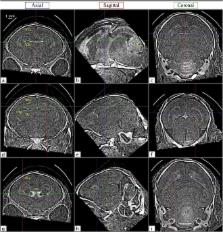
          Phase contrast X-ray imaging (PCXI) is an emerging imaging modality that has the potential to greatly improve radiography for medical imaging and materials analysis. PCXI makes it possible to visualise soft-tissue structures that are otherwise unresolved with conventional CT by rendering phase gradients in the X-ray wavefield visible. This can improve the contrast resolution of soft tissues structures, like the lungs and brain, by orders of magnitude. Phase retrieval suppresses noise, revealing weakly-attenuating soft tissue structures, however it does not remove the artefacts from the highly attenuating bone of the skull and from imperfections in the imaging system that can obscure those structures. The primary causes of these artefacts are investigated and a simple method to visualise the features they obstruct is proposed, which can easily be implemented for preclinical animal studies. We show that phase contrast X-ray CT (PCXI-CT) can resolve the soft tissues of the brain in situ without a need for contrast agents at a dose ~400 times lower than would be required by standard absorption contrast CT. We generalise a well-known phase retrieval algorithm for multiple-material samples specifically for CT, validate its use for brain CT, and demonstrate its high stability in the presence of noise.

          Related collections

          Most cited references38

          • Record: found
          • Abstract: not found
          • Article: not found

          Computerized transverse axial scanning (tomography). 1. Description of system.

            Bookmark
            • Record: found
            • Abstract: not found
            • Article: not found

            Phase-contrast imaging using polychromatic hard X-rays

              Bookmark
              • Record: found
              • Abstract: found
              • Article: not found

              The ASTRA Toolbox: A platform for advanced algorithm development in electron tomography.

              We present the ASTRA Toolbox as an open platform for 3D image reconstruction in tomography. Most of the software tools that are currently used in electron tomography offer limited flexibility with respect to the geometrical parameters of the acquisition model and the algorithms used for reconstruction. The ASTRA Toolbox provides an extensive set of fast and flexible building blocks that can be used to develop advanced reconstruction algorithms, effectively removing these limitations. We demonstrate this flexibility, the resulting reconstruction quality, and the computational efficiency of this toolbox by a series of experiments, based on experimental dual-axis tilt series.
                Bookmark

                Author and article information

                Contributors
                linda.croton@monash.edu
                Journal
                Sci Rep
                Sci Rep
                Scientific Reports
                Nature Publishing Group UK (London )
                2045-2322
                30 July 2018
                30 July 2018
                2018
                : 8
                : 11412
                Affiliations
                [1 ]ISNI 0000 0004 1936 7857, GRID grid.1002.3, School of Physics and Astronomy, , Monash University, Clayton, ; Victoria, 3800 Australia
                [2 ]ISNI 0000000123222966, GRID grid.6936.a, Chair of Biomedical Physics, Department of Physics, Munich School of Bioengineering, and Institute of Advanced Study, , Technische Universität München, ; 85748 Garching, Germany
                [3 ]GRID grid.452824.d, The Ritchie Centre, , Hudson Institute of Medical Research, Clayton, ; Victoria, 3800 Australia
                [4 ]ISNI 0000 0004 0422 0975, GRID grid.11485.39, Cancer Research UK, Angel, ; London, United Kingdom
                [5 ]ISNI 0000 0004 1936 7857, GRID grid.1002.3, Department of Obstetrics and Gynaecology, , Monash University, Clayton, ; Victoria, 3800 Australia
                [6 ]ISNI 0000 0001 2170 091X, GRID grid.410592.b, Japan Synchrotron Radiation Research Institute (JASRI/SPring-8), ; 1-1-1 Kouto, Sayo, Hyogo, 679-5198 Japan
                Article
                29841
                10.1038/s41598-018-29841-5
                6065359
                30061729
                89b86354-480c-4971-9396-5188794a1b2c
                © The Author(s) 2018

                Open Access This article is licensed under a Creative Commons Attribution 4.0 International License, which permits use, sharing, adaptation, distribution and reproduction in any medium or format, as long as you give appropriate credit to the original author(s) and the source, provide a link to the Creative Commons license, and indicate if changes were made. The images or other third party material in this article are included in the article’s Creative Commons license, unless indicated otherwise in a credit line to the material. If material is not included in the article’s Creative Commons license and your intended use is not permitted by statutory regulation or exceeds the permitted use, you will need to obtain permission directly from the copyright holder. To view a copy of this license, visit http://creativecommons.org/licenses/by/4.0/.

                History
                : 31 January 2018
                : 17 July 2018
                Funding
                Funded by: Australian Research Training Program
                Funded by: Veski Postdoctoral Research Fellowship
                Funded by: FundRef https://doi.org/10.13039/501100000923, Australian Research Council (ARC);
                Award ID: DP170103678
                Award Recipient :
                Categories
                Article
                Custom metadata
                © The Author(s) 2018

                Uncategorized
                Uncategorized

                Comments

                Comment on this article