0
views
0
recommends
+1 Recommend
0 collections
    0
    shares
      • Record: found
      • Abstract: found
      • Article: found
      Is Open Access

      Development of Cystoisospora felis in Cell Culture and in vitro Formation of Monozoic Tissue Cysts

      research-article

      Read this article at
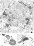
      Bookmark
          There is no author summary for this article yet. Authors can add summaries to their articles on ScienceOpen to make them more accessible to a non-specialist audience.

          Abstract

          Cystoisospora felis is a coccidian parasite commonly found in feces of domestic cats. Infection in cats occurs by ingestion of sporulated oocysts or consumption of rodents infected by the parasite. Scarce information is available about extraintestinal stages of C. felis in naturally infected intermediate hosts, as well as in cell culture. The aim of the current work was to investigate the development of C. felis in Vero cells (African green monkey kidney) and MDCK cells (Madin-Darby canine kidney). Cell monolayers were inoculated with mechanically released sporozoites of C. felis, and parasite growth was daily examined using light microscopy. After cell invasion, only parasitophorous vacuoles containing a single zoite were observed. Five days post-inoculation with sporozoites, unstained cell monolayers were evaluated by differential interference contrast (DIC), and also by Romanovsky stain using conventional light microscopy. Single zoites, each surrounded by a cyst wall, were observed by both methods. Multiplication by endodyogeny did not occur in any cell monolayer. Treatment of encysted parasites with HCl-pepsin for 15 min led to dissolution of the cyst wall and release of intact and motile zoites. To our knowledge, this is the first demonstration of in vitro production of monozoic tissue cysts of C. felis. As kittens commonly shed C. felis in their feces, oocysts are easily available for in vitro production of monozoic tissue cysts of the parasite. Development of C. felis in cell culture may be employed as a model on tissue cyst formation of Cystoisospora spp. and closely related coccidia.

          Related collections

          Most cited references21

          • Record: found
          • Abstract: found
          • Article: not found

          Neonatal Neospora caninum infection in dogs: isolation of the causative agent and experimental transmission.

          Neospora caninum infection was diagnosed in 5 young dogs from 2 litters with a common parentage. The pups were born healthy, but developed hind limb paresis 5 to 8 weeks after birth. The predominant lesions were polyradiculoneuritis and granulomatous polymyositis. Neospora caninum was seen microscopically in sections of naturally infected pups, and was isolated in cell cultures, mice, and dogs inoculated with infected canine tissues. Antibodies to N caninum were detected in sera of infected dogs by indirect fluorescent antibody test.
            Bookmark
            • Record: found
            • Abstract: found
            • Article: found
            Is Open Access

            Isospora suis in an Epithelial Cell Culture System – An In Vitro Model for Sexual Development in Coccidia

            Coccidian parasites are of major importance in animal production, public health and food safety. The most frequently used representative in basic research on this group is Toxoplasma gondii . Although this parasite is well investigated there is no adequate in vitro model for its sexual development available and knowledge on this important life cycle phase is therefore scarce. The use of Isospora suis , a sister taxon to T. gondii and the causative agent of piglet coccidiosis, could provide a solution for this. In the present study an in vitro model for neonatal porcine coccidiosis in cells representative for the in vivo situation in the piglet gut was developed and evaluated. The parasite development was investigated by light and transmission electron microscopy and optimum culture conditions were evaluated. Intestinal porcine epithelial cells (IPEC-J2) adequately representing the natural host cells supported the development of all endogenous life cycle stages of I . suis , including gametocytes and oocysts. A concentration of 5% fetal calf serum in the culture medium led to highest gametocyte densities on day 12 post infection. Low infection doses (≤1 sporozoite for 100 host cells) were best for oocyst and gametocyte development. The presented system can also be used for immunostaining with established antibodies developed against T. gondii (in our case, anti-TgIMC3 antibodies directed against the inner membrane complex 3). The complete life cycle of I . suis in a cell line representing the natural host cell type and species provides a unique model among coccidian parasites and can be used to address a wide range of topics, especially with regard to the sexual development of coccidia.
              Bookmark
              • Record: found
              • Abstract: found
              • Article: not found

              Comparison of a serum indirect fluorescent antibody test with two Western blot tests for the diagnosis of equine protozoal myeloencephalitis.

              A serum indirect fluorescent antibody test (IFAT) was compared with a Western blot (WB) and a modified Western blot (mWB) for diagnosis of equine protozoal myeloencephalitis (EPM). Using receiver-operating characteristic (ROC) analysis, the area under the curve of the IFAT was greater than the areaunder the curves of the WB and the mWB (P = 0.025 and P = 0.044, respectively). There was no statistically significant difference between the areas under the curves of the WBs (P > 0.05). On the basis of an arbitrarily chosen cut-off titer for a positive test result of 1:80 for the IFAT and interpreting weak positive WB results as positive test results, the sensitivities and 95% confidence intervals (CI) of all 3 tests were identical and equal to 88.9% (51.8-99.7%). The specificities and 95% CIs of the IFAT, WB, and mWB test were 100% (91-100%), 87.2% (72.6-95.7%), and 69.2% (52.4-83%), respectively. The overall accuracy of the IFAT was shown to be better than that of the WBs and, therefore, the test has potential for use in the diagnosis of EPM caused by Sarcocystis neurona.
                Bookmark

                Author and article information

                Contributors
                Journal
                Front Vet Sci
                Front Vet Sci
                Front. Vet. Sci.
                Frontiers in Veterinary Science
                Frontiers Media S.A.
                2297-1769
                16 October 2019
                2019
                : 6
                : 361
                Affiliations
                Department of Veterinary Anatomy, Pathology and Clinics, School of Veterinary Medicine and Animal Science, Federal University of Bahia , Salvador, Brazil
                Author notes

                Edited by: David Modrý, University of Veterinary and Pharmaceutical Sciences Brno, Czechia

                Reviewed by: David S. Lindsay, Virginia Tech, United States; Rafael Calero-Bernal, Complutense University of Madrid, Spain

                *Correspondence: Luís F. P. Gondim pita@ 123456ufba.br

                This article was submitted to Parasitology, a section of the journal Frontiers in Veterinary Science

                Article
                10.3389/fvets.2019.00361
                6805693
                8a7173f5-9686-4153-8e60-0aaa616735f4
                Copyright © 2019 Ferreira, Borges-Silva, de Jesus and Gondim.

                This is an open-access article distributed under the terms of the Creative Commons Attribution License (CC BY). The use, distribution or reproduction in other forums is permitted, provided the original author(s) and the copyright owner(s) are credited and that the original publication in this journal is cited, in accordance with accepted academic practice. No use, distribution or reproduction is permitted which does not comply with these terms.

                History
                : 05 July 2019
                : 01 October 2019
                Page count
                Figures: 2, Tables: 1, Equations: 0, References: 21, Pages: 5, Words: 3693
                Funding
                Funded by: Conselho Nacional de Desenvolvimento Científico e Tecnológico 10.13039/501100003593
                Funded by: Coordenação de Aperfeiçoamento de Pessoal de Nível Superior 10.13039/501100002322
                Categories
                Veterinary Science
                Original Research

                isospora felis,cystoisospora sp.,tissue cyst,sporozoite,oocyst

                Comments

                Comment on this article