32
views
0
recommends
+1 Recommend
0 collections
    0
    shares
      • Record: found
      • Abstract: found
      • Article: found
      Is Open Access

      Protective but Not Anticonvulsant Effects of Ghrelin and JMV-1843 in the Pilocarpine Model of Status epilepticus

      research-article

      Read this article at

      Bookmark
          There is no author summary for this article yet. Authors can add summaries to their articles on ScienceOpen to make them more accessible to a non-specialist audience.

          Abstract

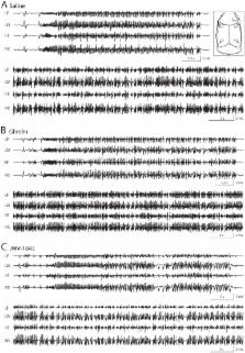
          In models of status epilepticus ghrelin displays neuroprotective effects mediated by the growth hormone secretagogue-receptor 1a (GHS-R 1a). This activity may be explained by anticonvulsant properties that, however, are controversial. We further investigated neuroprotection and the effects on seizures by comparing ghrelin with a more effective GHS-R 1a agonist, JMV-1843. Rats were treated either with ghrelin, JMV-1843 or saline 10 min before pilocarpine, which was used to induce status epilepticus. Status epilepticus, developed in all rats, was attenuated by diazepam. No differences were observed among the various groups in the characteristics of pilocarpine-induced seizures. In saline group the area of lesion, characterized by lack of glial fibrillary acidic protein immunoreactivity, was of 0.45±0.07 mm 2 in the hippocampal stratum lacunosum-moleculare, and was accompanied by upregulation of laminin immunostaining, and by increased endothelin-1 expression. Both ghrelin ( P<0.05) and JMV-1843 ( P<0.01) were able to reduce the area of loss in glial fibrillary acidic protein immunostaining. In addition, JMV-1843 counteracted ( P<0.05) the changes in laminin and endothelin-1 expression, both increased in ghrelin-treated rats. JMV-1843 was able to ameliorate neuronal survival in the hilus of dentate gyrus and medial entorhinal cortex layer III ( P<0.05 vs saline and ghrelin groups). These results demonstrate diverse protective effects of growth hormone secretagogues in rats exposed to status epilepticus.

          Related collections

          Most cited references48

          • Record: found
          • Abstract: found
          • Article: not found

          The pilocarpine model of temporal lobe epilepsy

          Understanding the pathophysiogenesis of temporal lobe epilepsy (TLE) largely rests on the use of models of status epilepticus (SE), as in the case of the pilocarpine model. The main features of TLE are: (i) epileptic foci in the limbic system; (ii) an “initial precipitating injury”; (iii) the so-called “latent period”; and (iv) the presence of hippocampal sclerosis leading to reorganization of neuronal networks. Many of these characteristics can be reproduced in rodents by systemic injection of pilocarpine; in this animal model, SE is followed by a latent period and later by the appearance of spontaneous recurrent seizures (SRSs). These processes are, however, influenced by experimental conditions such as rodent species, strain, gender, age, doses and routes of pilocarpine administration, as well as combinations with other drugs administered before and/or after SE. In the attempt to limit these sources of variability, we evaluated the methodological procedures used by several investigators in the pilocarpine model; in particular, we have focused on the behavioural, electrophysiological and histopathological findings obtained with different protocols. We addressed the various experimental approaches published to date, by comparing mortality rates, onset of SRSs, neuronal damage, and network reorganization. Based on the evidence reviewed here, we propose that the pilocarpine model can be a valuable tool to investigate the mechanisms involved in TLE, and even more so when standardized to reduce mortality at the time of pilocarpine injection, differences in latent period duration, variability in the lesion extent, and SRS frequency.
            Bookmark
            • Record: found
            • Abstract: found
            • Article: not found

            Fluoro-Jade B: a high affinity fluorescent marker for the localization of neuronal degeneration.

            Fluoro-Jade B, like its predecessor Fluoro-Jade, is an anionic fluorescein derivative useful for the histological staining of neurons undergoing degeneration. However, Fluoro-Jade B has an even greater specific affinity for degenerating neurons. This notion is supported by the conspicuous staining of degenerating neuronal elements with minimal background staining. This improved signal-to-noise ratio means that fine neuronal processes including distal dendrites, axons and axon terminals can be more readily detected and documented. Although the staining time and dye concentration are reduced, the method is as rapid, simple and reliable as the original Fluoro-Jade technique. Like Fluoro-Jade, Fluoro-Jade B is compatible with a number of other labeling procedures including immunofluorescent and fluorescent Nissl techniques.
              Bookmark
              • Record: found
              • Abstract: found
              • Article: not found

              Ghrelin and des-acyl ghrelin inhibit cell death in cardiomyocytes and endothelial cells through ERK1/2 and PI 3-kinase/AKT

              Ghrelin is an acyl-peptide gastric hormone acting on the pituitary and hypothalamus to stimulate growth hormone (GH) release, adiposity, and appetite. Ghrelin endocrine activities are entirely dependent on its acylation and are mediated by GH secretagogue (GHS) receptor (GHSR)-1a, a G protein–coupled receptor mostly expressed in the pituitary and hypothalamus, previously identified as the receptor for a group of synthetic molecules featuring GH secretagogue (GHS) activity. Des-acyl ghrelin, which is far more abundant than ghrelin, does not bind GHSR-1a, is devoid of any endocrine activity, and its function is currently unknown. Ghrelin, which is expressed in heart, albeit at a much lower level than in the stomach, also exerts a cardio protective effect through an unknown mechanism, independent of GH release. Here we show that both ghrelin and des-acyl ghrelin inhibit apoptosis of primary adult and H9c2 cardiomyocytes and endothelial cells in vitro through activation of extracellular signal–regulated kinase-1/2 and Akt serine kinases. In addition, ghrelin and des-acyl ghrelin recognize common high affinity binding sites on H9c2 cardiomyocytes, which do not express GHSR-1a. Finally, both MK-0677 and hexarelin, a nonpeptidyl and a peptidyl synthetic GHS, respectively, recognize the common ghrelin and des-acyl ghrelin binding sites, inhibit cell death, and activate MAPK and Akt. These findings provide the first evidence that, independent of its acylation, ghrelin gene product may act as a survival factor directly on the cardiovascular system through binding to a novel, yet to be identified receptor, which is distinct from GHSR-1a.
                Bookmark

                Author and article information

                Contributors
                Role: Editor
                Journal
                PLoS One
                PLoS ONE
                plos
                plosone
                PLoS ONE
                Public Library of Science (San Francisco, USA )
                1932-6203
                2013
                28 August 2013
                : 8
                : 8
                : e72716
                Affiliations
                [1 ]Department of Biomedical, Metabolic and Neural Sciences, University of Modena and Reggio Emilia, Modena, Italy
                [2 ]Department of Health Sciences, University of Milano-Bicocca, Monza, Italy
                [3 ]Department of Neurosciences, NOCSAE Hospital, Modena, Italy
                Beijing Institute of Radiation Medicine, China
                Author notes

                Competing Interests: The authors have declared that no competing interests exist.

                Conceived and designed the experiments: EB VL AT GB. Performed the experiments: CL GC JV FG. Analyzed the data: CL GC GB. Contributed reagents/materials/analysis tools: EB VL AT GB. Wrote the paper: GC JV AT GB.

                Article
                PONE-D-13-10146
                10.1371/journal.pone.0072716
                3755992
                24015271
                90346915-bbdc-48e9-afff-c8f197b964a4
                Copyright @ 2013

                This is an open-access article distributed under the terms of the Creative Commons Attribution License, which permits unrestricted use, distribution, and reproduction in any medium, provided the original author and source are credited.

                History
                : 11 March 2013
                : 15 July 2013
                Page count
                Pages: 13
                Funding
                This study was supported by the Ministry of Health (grant RF-2010-2309921 to GB; https://www.salute.gov.it/ricercaSanitaria/ricercaSanitaria.jsp), the University of Milano-Bicocca (Fondo di Ateneo per la Ricerca FAR, to AT and VL; http://www.unimib.it/go/101/Home/Italiano) and the Italian Ministry of Education, University and Research (“Rientro Cervelli” project 17DZE8RZEA to GC; http://sito.cineca.it/). The funders had no role in study design, data collection and analysis, decision to publish, or preparation of the manuscript.
                Categories
                Research Article
                Biology
                Anatomy and Physiology
                Electrophysiology
                Endocrine System
                Neurological System
                Neuroscience
                Behavioral Neuroscience
                Cellular Neuroscience
                Neurobiology of Disease and Regeneration
                Neurochemistry
                Medicine
                Neurology
                Cerebrovascular Diseases
                Epilepsy
                Neuropharmacology

                Uncategorized
                Uncategorized

                Comments

                Comment on this article