23
views
0
recommends
+1 Recommend
0 collections
    0
    shares
      • Record: found
      • Abstract: found
      • Article: found
      Is Open Access

      Perineuronal Nets and Their Role in Synaptic Homeostasis

      review-article

      Read this article at

      Bookmark
          There is no author summary for this article yet. Authors can add summaries to their articles on ScienceOpen to make them more accessible to a non-specialist audience.

          Abstract

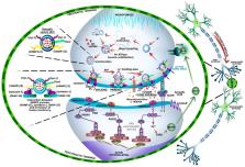
          Extracellular matrix (ECM) molecules that are released by neurons and glial cells form perineuronal nets (PNNs) and modulate many neuronal and glial functions. PNNs, whose structure is still not known in detail, surround cell bodies and dendrites, which leaves free space for synapses to come into contact. A reduction in the expression of many neuronal ECM components adversely affects processes that are associated with synaptic plasticity, learning, and memory. At the same time, increased ECM activity, e.g., as a result of astrogliosis following brain damage or in neuroinflammation, can also have harmful consequences. The therapeutic use of enzymes to attenuate elevated neuronal ECM expression after injury or in Alzheimer’s disease has proven to be beneficial by promoting axon growth and increasing synaptic plasticity. Yet, severe impairment of ECM function can also lead to neurodegeneration. Thus, it appears that to ensure healthy neuronal function a delicate balance of ECM components must be maintained. In this paper we review the structure of PNNs and their components, such as hyaluronan, proteoglycans, core proteins, chondroitin sulphate proteoglycans, tenascins, and Hapln proteins. We also characterize the role of ECM in the functioning of the blood-brain barrier, neuronal communication, as well as the participation of PNNs in synaptic plasticity and some clinical aspects of perineuronal net impairment. Furthermore, we discuss the participation of PNNs in brain signaling. Understanding the molecular foundations of the ways that PNNs participate in brain signaling and synaptic plasticity, as well as how they change in physiological and pathological conditions, may help in the development of new therapies for many degenerative and inflammatory diseases of the brain.

          Related collections

          Most cited references94

          • Record: found
          • Abstract: found
          • Article: not found

          Membrane fusion: grappling with SNARE and SM proteins.

          The two universally required components of the intracellular membrane fusion machinery, SNARE and SM (Sec1/Munc18-like) proteins, play complementary roles in fusion. Vesicular and target membrane-localized SNARE proteins zipper up into an alpha-helical bundle that pulls the two membranes tightly together to exert the force required for fusion. SM proteins, shaped like clasps, bind to trans-SNARE complexes to direct their fusogenic action. Individual fusion reactions are executed by distinct combinations of SNARE and SM proteins to ensure specificity, and are controlled by regulators that embed the SM-SNARE fusion machinery into a physiological context. This regulation is spectacularly apparent in the exquisite speed and precision of synaptic exocytosis, where synaptotagmin (the calcium-ion sensor for fusion) cooperates with complexin (the clamp activator) to control the precisely timed release of neurotransmitters that initiates synaptic transmission and underlies brain function.
            Bookmark
            • Record: found
            • Abstract: found
            • Article: not found

            Perineuronal nets protect fear memories from erasure.

            In adult animals, fear conditioning induces a permanent memory that is resilient to erasure by extinction. In contrast, during early postnatal development, extinction of conditioned fear leads to memory erasure, suggesting that fear memories are actively protected in adults. We show here that this protection is conferred by extracellular matrix chondroitin sulfate proteoglycans (CSPGs) in the amygdala. The organization of CSPGs into perineuronal nets (PNNs) coincided with the developmental switch in fear memory resilience. In adults, degradation of PNNs by chondroitinase ABC specifically rendered subsequently acquired fear memories susceptible to erasure. This result indicates that intact PNNs mediate the formation of erasure-resistant fear memories and identifies a molecular mechanism closing a postnatal critical period during which traumatic memories can be erased by extinction.
              Bookmark
              • Record: found
              • Abstract: found
              • Article: not found

              The postsynaptic organization of synapses.

              The postsynaptic side of the synapse is specialized to receive the neurotransmitter signal released from the presynaptic terminal and transduce it into electrical and biochemical changes in the postsynaptic cell. The cardinal functional components of the postsynaptic specialization of excitatory and inhibitory synapses are the ionotropic receptors (ligand-gated channels) for glutamate and γ-aminobutyric acid (GABA), respectively. These receptor channels are concentrated at the postsynaptic membrane and embedded in a dense and rich protein network comprised of anchoring and scaffolding molecules, signaling enzymes, cytoskeletal components, as well as other membrane proteins. Excitatory and inhibitory postsynaptic specializations are quite different in molecular organization. The postsynaptic density of excitatory synapses is especially complex and dynamic in composition and regulation; it contains hundreds of different proteins, many of which are required for cognitive function and implicated in psychiatric illness.
                Bookmark

                Author and article information

                Journal
                Int J Mol Sci
                Int J Mol Sci
                ijms
                International Journal of Molecular Sciences
                MDPI
                1422-0067
                22 August 2019
                September 2019
                : 20
                : 17
                : 4108
                Affiliations
                [1 ]Department of Functional Diagnostics and Physical Medicine, Pomeranian Medical University in Szczecin, Żołnierska 54 Str., 71-210 Szczecin, Poland
                [2 ]Department of Cellular Signalling, Mossakowski Medical Research Centre, Polish Academy of Sciences, Pawińskiego 5 Str., 02-106 Warsaw, Poland
                [3 ]Department of Anaesthesiology and Intensive Care, Pomeranian Medical University in Szczecin, 71-252 Szczecin, Poland
                [4 ]Institute of Philosophy, University of Szczecin, Krakowska 71-79 Str., 71-017 Szczecin, Poland
                [5 ]Department of Neurosurgery and Pediatric Neurosurgery, Department of Applied Neurocognitivistics, Pomeranian Medical University in Szczecin, 71-252 Szczecin, Poland
                [6 ]Department of Human Nutrition and Metabolomics, Department of Medical Chemistry, Pomeranian Medical University in Szczecin, Broniewskiego 24 Str., 71-252 Szczecin, Poland
                Author notes
                [* ]Correspondence: e-mail: anna.lubkowska@ 123456pum.edu.pl
                Author information
                https://orcid.org/0000-0001-8823-0473
                https://orcid.org/0000-0002-5378-5409
                Article
                ijms-20-04108
                10.3390/ijms20174108
                6747153
                31443560
                90900bc7-ca16-435b-aa85-86c46506f69f
                © 2019 by the authors.

                Licensee MDPI, Basel, Switzerland. This article is an open access article distributed under the terms and conditions of the Creative Commons Attribution (CC BY) license ( http://creativecommons.org/licenses/by/4.0/).

                History
                : 07 July 2019
                : 16 August 2019
                Categories
                Review

                Molecular biology
                perineuronal nets (pnns),extracellular matrix (ecm),synaptogenesis,neuronal communication

                Comments

                Comment on this article