18
views
0
recommends
+1 Recommend
0 collections
    0
    shares
      • Record: found
      • Abstract: found
      • Article: not found

      Temporal Evolution of Cortical Ensembles Promoting Remote Memory Retrieval

      research-article

      Read this article at

      ScienceOpenPublisherPMC
      Bookmark
          There is no author summary for this article yet. Authors can add summaries to their articles on ScienceOpen to make them more accessible to a non-specialist audience.

          Abstract

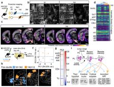
          Memories of fearful events can last a lifetime. The prelimbic (PL) subregion of prefrontal cortex plays a critical role in fear memory retrieval over time. Most studies have focused on acquisition, consolidation, and retrieval of recent memories, but much less is known about the neural mechanisms of remote memory. Using a new knock-in mouse for activity-dependent genetic labeling (TRAP2), we demonstrate that neuronal ensembles in PL are dynamic. PL neurons TRAPed during later memory retrievals are more likely to be reactivated and make larger behavioral contributions to remote memory retrieval compared to those TRAPed during learning or early memory retrieval. PL activity during learning is required to initiate this time-dependent reorganization in PL ensembles underlying memory retrieval. Finally, while neurons TRAPed during earlier and later retrievals have similar broad projections throughout the brain, PL neurons TRAPed later have a stronger functional recruitment of cortical targets.

          Related collections

          Most cited references33

          • Record: found
          • Abstract: found
          • Article: not found

          Interplay of hippocampus and prefrontal cortex in memory.

          Recent studies on the hippocampus and the prefrontal cortex have considerably advanced our understanding of the distinct roles of these brain areas in the encoding and retrieval of memories, and of how they interact in the prolonged process by which new memories are consolidated into our permanent storehouse of knowledge. These studies have led to a new model of how the hippocampus forms and replays memories and how the prefrontal cortex engages representations of the meaningful contexts in which related memories occur, as well as how these areas interact during memory retrieval. Furthermore, they have provided new insights into how interactions between the hippocampus and prefrontal cortex support the assimilation of new memories into pre-existing networks of knowledge, called schemas, and how schemas are modified in this process as the foundation of memory consolidation. Copyright © 2013 Elsevier Ltd. All rights reserved.
            Bookmark
            • Record: found
            • Abstract: found
            • Article: not found

            Long-term dynamics of CA1 hippocampal place codes

            Via Ca2+-imaging in freely behaving mice that repeatedly explored a familiar environment, we tracked thousands of CA1 pyramidal cells' place fields over weeks. Place coding was dynamic, for each day the ensemble representation of this environment involved a unique subset of cells. Yet, cells within the ∼15–25% overlap between any two of these subsets retained the same place fields, which sufficed to preserve an accurate spatial representation across weeks.
              Bookmark
              • Record: found
              • Abstract: found
              • Article: not found

              Dissociable roles of prelimbic and infralimbic cortices, ventral hippocampus, and basolateral amygdala in the expression and extinction of conditioned fear.

              Current models of conditioned fear expression and extinction involve the basolateral amygdala (BLA), ventral medial prefrontal cortex (vmPFC), and the hippocampus (HPC). There is some disagreement with respect to the specific roles of these structures, perhaps due to subregional differences within each area. For example, growing evidence suggests that infralimbic (IL) and prelimbic (PL) subregions of vmPFC have opposite influences on fear expression. Moreover, it is the ventral HPC (vHPC), rather than the dorsal HPC, that projects to vmPFC and BLA. To help determine regional specificity, we used small doses of the GABA(A) agonist muscimol to selectively inactivate IL, PL, BLA, or vHPC in an auditory fear conditioning and extinction paradigm. Infusions were performed prior to extinction training, allowing us to assess the effects on both fear expression and subsequent extinction memory. Inactivation of IL had no effect on fear expression, but impaired the within-session acquisition of extinction as well as extinction memory. In contrast, inactivation of PL impaired fear expression, but had no effect on extinction memory. Inactivation of the BLA or vHPC impaired both fear expression and extinction memory. Post-extinction inactivations had no effect in any structure. We suggest a model in which amygdala-dependent fear expression is modulated by inputs from PL and vHPC, whereas extinction memory requires extinction-induced plasticity in IL, BLA, and/or vHPC.
                Bookmark

                Author and article information

                Journal
                9809671
                21092
                Nat Neurosci
                Nat. Neurosci.
                Nature neuroscience
                1097-6256
                1546-1726
                28 January 2019
                28 January 2019
                March 2019
                28 July 2019
                : 22
                : 3
                : 460-469
                Affiliations
                [1 ]Department of Biology, Stanford University, Stanford, California, USA
                [2 ]Howard Hughes Medical Institute, Stanford University, Stanford, California, USA
                [3 ]Neurosciences Program, Stanford University, Stanford, California, USA
                Author notes

                Author Contributions L.A.D. and L.L. designed experiments. L.A.D. and C.J.G. generated the TRAP2 targeting construct. L.A.D. characterized the TRAP2 mouse. L.A.D. and C.D.L. performed behavior assays. L.A.D. and L.F. performed histology and confocal imaging. L.A.D., C.D.L., and L.F. analyzed the data, W.E.A. analyzed iDISCO+-generated data sets. E.L.A. (with support from M.T.-L.) advised and provided training in the iDISCO+ and ClearMap methods. D.F. wrote software and advised for quantitative whole-brain axon analysis. L.A.D. and L.L. wrote the manuscript.

                [* ]Corresponding authors. ldenardo@ 123456ucla.edu (L.A.D.), lluo@ 123456stanford.edu (L.L.)
                Additional information Correspondence and requests for materials should be addressed to L.A.D. or L.L..
                Article
                NIHMS1515987
                10.1038/s41593-018-0318-7
                6387639
                30692687
                90a335f1-3aa9-4e6d-88f2-d998bb4d244b

                Users may view, print, copy, and download text and data-mine the content in such documents, for the purposes of academic research, subject always to the full Conditions of use: http://www.nature.com/authors/editorial_policies/license.html#terms

                History
                Categories
                Article

                Neurosciences
                Neurosciences

                Comments

                Comment on this article