57
views
0
recommends
+1 Recommend
0 collections
    0
    shares
      • Record: found
      • Abstract: found
      • Article: found
      Is Open Access

      Microdomains of muscarinic acetylcholine and Ins(1,4,5) P 3 receptors create ‘Ins(1,4,5) P 3 junctions’ and sites of Ca 2+ wave initiation in smooth muscle

      research-article

      Read this article at

      Bookmark
          There is no author summary for this article yet. Authors can add summaries to their articles on ScienceOpen to make them more accessible to a non-specialist audience.

          Summary

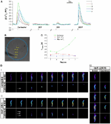
          Increases in cytosolic Ca 2+ concentration ([Ca 2+] c) mediated by inositol (1,4,5)-trisphosphate [Ins(1,4,5) P 3, hereafter InsP 3] regulate activities that include division, contraction and cell death. InsP 3-evoked Ca 2+ release often begins at a single site, then regeneratively propagates through the cell as a Ca 2+ wave. The Ca 2+ wave consistently begins at the same site on successive activations. Here, we address the mechanisms that determine the Ca 2+ wave initiation site in intestinal smooth muscle cells. Neither an increased sensitivity of InsP 3 receptors (InsP 3R) to InsP 3 nor regional clustering of muscarinic receptors (mAChR3) or InsP 3R1 explained the selection of an initiation site. However, examination of the overlap of mAChR3 and InsP 3R1 localisation, by centre of mass analysis, revealed that there was a small percentage (∼10%) of sites that showed colocalisation. Indeed, the extent of colocalisation was greatest at the Ca 2+ wave initiation site. The initiation site might arise from a selective delivery of InsP 3 from mAChR3 activity to particular InsP 3Rs to generate faster local [Ca 2+] c increases at sites of colocalisation. In support of this hypothesis, a localised subthreshold ‘priming’ InsP 3 concentration applied rapidly, but at regions distant from the initiation site, shifted the wave to the site of the priming. Conversely, when the Ca 2+ rise at the initiation site was rapidly and selectively attenuated, the Ca 2+ wave again shifted and initiated at a new site. These results indicate that Ca 2+ waves initiate where there is a structural and functional coupling of mAChR3 and InsP 3R1, which generates junctions in which InsP 3 acts as a highly localised signal by being rapidly and selectively delivered to InsP 3R1.

          Related collections

          Most cited references42

          • Record: found
          • Abstract: found
          • Article: not found

          Subtype-specific and ER lumenal environment-dependent regulation of inositol 1,4,5-trisphosphate receptor type 1 by ERp44.

          Inositol 1,4,5-trisphosphate receptors (IP(3)Rs) are intracellular channel proteins that mediate Ca(2+) release from the endoplasmic reticulum (ER) and are involved in many biological processes and diseases. IP(3)Rs are differentially regulated by a variety of cytosolic proteins, but their regulation by ER lumenal protein(s) remains largely unexplored. In this study, we found that ERp44, an ER lumenal protein of the thioredoxin family, directly interacts with the third lumenal loop of IP(3)R type 1 (IP(3)R1) and that the interaction is dependent on pH, Ca(2+) concentration, and redox state: the presence of free cysteine residues in the loop is required. Ca(2+)-imaging experiments and single-channel recording of IP(3)R1 activity with a planar lipid bilayer system demonstrated that IP(3)R1 is directly inhibited by ERp44. Thus, ERp44 senses the environment in the ER lumen and modulates IP(3)R1 activity accordingly, which should in turn contribute to regulating both intralumenal conditions and the complex patterns of cytosolic Ca(2+) concentrations.
            Bookmark
            • Record: found
            • Abstract: found
            • Article: not found

            Local and global cytosolic Ca2+ oscillations in exocrine cells evoked by agonists and inositol trisphosphate.

            Submaximal stimulation with agonists generating inositol 1,4,5-trisphosphate (IP3) evokes cytosolic Ca2+ oscillations in many different cell types. In general, each Ca2+ rise is initiated from a specific region near the plasma membrane and then spreads as a wave throughout the cell. We now demonstrate that low (physiological) agonist concentrations evoke local cytosolic Ca2+ spikes in the secretory pole of single mouse pancreatic acinar cells that are particularly sensitive to blockade by the IP3 receptor antagonist heparin. These spikes can occur alone or repetitively or can precede longer lasting Ca2+ signals that spread throughout the cell. Intracellular IP3 application mimics these agonist actions. The short-lasting local Ca2+ spikes provide an economical signaling mechanism and are of physiological significance since they activate Ca(2+)-dependent Cl- and cation currents important for control of fluid secretion.
              Bookmark
              • Record: found
              • Abstract: found
              • Article: not found

              Imaging the quantal substructure of single IP3R channel activity during Ca2+ puffs in intact mammalian cells.

              The spatiotemporal patterning of Ca(2+) signals regulates numerous cellular functions, and is determined by the functional properties and spatial clustering of inositol trisphosphate receptor (IP(3)R) Ca(2+) release channels in the endoplasmic reticulum membrane. However, studies at the single-channel level have been hampered because IP(3)Rs are inaccessible to patch-clamp recording in intact cells, and because excised organelle and bilayer reconstitution systems disrupt the Ca(2+)-induced Ca(2+) release (CICR) process that mediates channel-channel coordination. We introduce here the use of total internal reflection fluorescence microscopy to image single-channel Ca(2+) flux through individual and clustered IP(3)Rs in intact mammalian cells. This enables a quantal dissection of the local calcium puffs that constitute building blocks of cellular Ca(2+) signals, revealing stochastic recruitment of, on average, approximately 6 active IP(3)Rs clustered within <500 nm. Channel openings are rapidly ( approximately 10 ms) recruited by opening of an initial trigger channel, and a similarly rapid inhibitory process terminates puffs despite local [Ca(2+)] elevation that would otherwise sustain Ca(2+)-induced Ca(2+) release indefinitely. Minimally invasive, nano-scale Ca(2+) imaging provides a powerful tool for the functional study of intracellular Ca(2+) release channels while maintaining the native architecture and dynamic interactions essential for discrete and selective cell signaling.
                Bookmark

                Author and article information

                Journal
                J Cell Sci
                J. Cell. Sci
                joces
                jcs
                Journal of Cell Science
                The Company of Biologists (Bidder Building, 140 Cowley Road, Cambridge, CB4 0DL, UK )
                0021-9533
                1477-9137
                15 November 2012
                : 125
                : 22
                : 5315-5328
                Affiliations
                [1]Strathclyde Institute of Pharmacy and Biomedical Sciences, University of Strathclyde , The Arbuthnott Building, 161 Cathedral Street, Glasgow G4 0RE, UK
                Author notes
                [* ]Author for correspondence ( john.mccarron@ 123456strath.ac.uk )
                Article
                10.1242/jcs.105163
                3561854
                22946060
                916eab63-940d-4a06-be71-3b614608b422
                © 2012. Published by The Company of Biologists Ltd

                This is an Open Access article distributed under the terms of the Creative Commons Attribution Non-Commercial Share Alike License ( http://creativecommons.org/licenses/by-nc-sa/3.0/), which permits unrestricted non-commercial use, distribution and reproduction in any medium provided that the original work is properly cited and all further distributions of the work or adaptation are subject to the same Creative Commons License terms.

                History
                : 25 July 2012
                Categories
                Research Article

                Cell biology
                insp3,ca2+ waves,smooth muscle
                Cell biology
                insp3, ca2+ waves, smooth muscle

                Comments

                Comment on this article