12
views
0
recommends
+1 Recommend
1 collections
    0
    shares

      Clinical Interventions in Aging (submit here)

      This international, peer-reviewed Open Access journal by Dove Medical Press focuses on prevention and treatment of diseases in people over 65 years of age. Sign up for email alerts here.

      36,334 Monthly downloads/views I 3.829 Impact Factor I 7.4 CiteScore I 1.83 Source Normalized Impact per Paper (SNIP) I 1.044 Scimago Journal & Country Rank (SJR)

      • Record: found
      • Abstract: found
      • Article: found
      Is Open Access

      Thalamic Atrophy Plays a Crucial Role in the Effect of Asymptomatic Carotid Stenosis on Cognitive Impairment

      research-article

      Read this article at

      ScienceOpenPublisherPMC
      Bookmark
          There is no author summary for this article yet. Authors can add summaries to their articles on ScienceOpen to make them more accessible to a non-specialist audience.

          Abstract

          Purpose

          Our objectives were to assess the abnormalities of subcortical nuclei by combining volume and shape analyses and potential association with cognitive impairment.

          Patients and Methods

          Twenty-nine patients with severe ACS of the unilateral internal carotid artery and 31 controls were enrolled between January 2017 to August 2018. All participants underwent a comprehensive neuropsychological evaluation, blood lipid biochemical measurements, and structural magnetic resonance imaging (MRI) to measure subcortical volumes and sub-regional shape deformations. Basic statistics, correction for multiple comparisons. Seventeen ACS patients underwent carotid endarterectomy (CEA) within one week after baseline measurements, cognitive assessments and MRI scans were repeated 6 months after CEA.

          Results

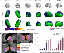
          The ACS patients had higher apolipoprotein B/apolipoprotein A1 (ApoB/ApoA1) ratio and worse performance in all cognitive domains than controls. Moreover, the ACS patients showed more profound thalamic atrophy assessed by shape and volume analysis, especially in the medial dorsal thalamus. No significant differences were found in other subcortical nuclei after multiple comparisons correction. At baseline, thalamic atrophy correlated with cognitive impairment and ApoB/ApoA1 ratio. Furthermore, mediation analysis at baseline showed that the association of carotid intima-media thickness with executive functioning was mediated by thalamic volume. After CEA, cognitive improvement and increase in the bilateral medial dorsal thalamic volume were observed.

          Conclusion

          Our study identified the distinct atrophy of subcortical nuclei and their association with cognition in patients with ACS. Assessments of the thalamus by volumetric and shape analysis may provide an early marker for cerebral ischemia and reperfusion after CEA.

          Most cited references33

          • Record: found
          • Abstract: found
          • Article: not found

          FSL.

          FSL (the FMRIB Software Library) is a comprehensive library of analysis tools for functional, structural and diffusion MRI brain imaging data, written mainly by members of the Analysis Group, FMRIB, Oxford. For this NeuroImage special issue on "20 years of fMRI" we have been asked to write about the history, developments and current status of FSL. We also include some descriptions of parts of FSL that are not well covered in the existing literature. We hope that some of this content might be of interest to users of FSL, and also maybe to new research groups considering creating, releasing and supporting new software packages for brain image analysis. Copyright © 2011 Elsevier Inc. All rights reserved.
            Bookmark
            • Record: found
            • Abstract: found
            • Article: found

            Dementia prevention, intervention, and care

              Bookmark
              • Record: found
              • Abstract: found
              • Article: not found

              MR signal abnormalities at 1.5 T in Alzheimer's dementia and normal aging.

              The type, frequency, and extent of MR signal abnormalities in Alzheimer's disease and normal aging are a subject of controversy. With a 1.5-MR unit we studied 12 Alzheimer patients, four subjects suffering from multiinfarct dementia and nine age-matched controls. Punctate or early confluent high-signal abnormalities in the deep white matter, noted in 60% of both Alzheimer patients and controls, were unrelated to the presence of hypertension or other vascular risk factors. A significant number of Alzheimer patients exhibited a more extensive smooth "halo" of periventricular hyperintensity when compared with controls (p = .024). Widespread deep white-matter hyperintensity (two patients) and extensive, irregular periventricular hyperintensity (three patients) were seen in multiinfarct dementia. Areas of high signal intensity affecting hippocampal and sylvian cortex were also present in five Alzheimer and two multiinfarct dementia patients, but absent in controls. Discrete, small foci of deep white-matter hyperintensity are not characteristic of Alzheimer's disease nor do they appear to imply a vascular cause for the dementing illness. The frequently observed "halo" of periventricular hyperintensity in Alzheimer's disease may be of diagnostic importance. High-signal abnormalities in specific cortical regions are likely to reflect disease processes localized to those structures.
                Bookmark

                Author and article information

                Journal
                Clin Interv Aging
                Clin Interv Aging
                cia
                clinintag
                Clinical Interventions in Aging
                Dove
                1176-9092
                1178-1998
                06 November 2020
                2020
                : 15
                : 2083-2094
                Affiliations
                [1 ]Department of Radiology, Drum Tower Hospital, The Affiliated Hospital of Nanjing University Medical School , Nanjing, People’s Republic of China
                [2 ]Department of Vascular and Interventional Radiology, Wujin People’s Hospital Affiliated to Jiangsu University , Changzhou, People’s Republic of China
                [3 ]Department of Neurology, Memory Clinic, 2nd Faculty of Medicine, Charles University and Motol University Hospital , Prague, Czech Republic
                [4 ]Columbia University Vagelos College of Physicians and Surgeons , New York, NY, USA
                [5 ]Department of Vascular Surgery, Drum Tower Hospital, The Affiliated Hospital of Nanjing University Medical School , Nanjing, People’s Republic of China
                Author notes
                Correspondence: Bing Zhang Department of Radiology, Drum Tower Hospital, The Affiliated Hospital of Nanjing University Medical School , No. 321 Zhongshan Road, Nanjing210008, People’s Republic of ChinaTel +86 15851803070 Email zhangbing_nanjing@nju.edu.cn
                Tong Qiao Department of Vascular Surgery, Drum Tower Hospital, The Affiliated Hospital of Nanjing University Medical School , No. 321 Zhongshan Road, Nanjing210008, People’s Republic of ChinaTel +86 15951985338 Email qiaotongmail@aliyun.com
                Author information
                http://orcid.org/0000-0001-5400-7791
                http://orcid.org/0000-0002-1080-3726
                Article
                273185
                10.2147/CIA.S273185
                7654547
                91ceb1e2-c83b-4ece-97e8-040b16e90415
                © 2020 Zhang et al.

                This work is published and licensed by Dove Medical Press Limited. The full terms of this license are available at https://www.dovepress.com/terms.php and incorporate the Creative Commons Attribution – Non Commercial (unported, v3.0) License ( http://creativecommons.org/licenses/by-nc/3.0/). By accessing the work you hereby accept the Terms. Non-commercial uses of the work are permitted without any further permission from Dove Medical Press Limited, provided the work is properly attributed. For permission for commercial use of this work, please see paragraphs 4.2 and 5 of our Terms ( https://www.dovepress.com/terms.php).

                History
                : 27 July 2020
                : 21 October 2020
                Page count
                Figures: 3, Tables: 12, References: 33, Pages: 12
                Funding
                Funded by: the National Natural Science Foundation of China;
                Funded by: the International Brain Research Organization and International Society for Neurochemistry (IBRO-ISN) return home fellowship 2018;
                This study was supported by the National Natural Science Foundation of China (grant number 81370387 and 81720108022). Zuzana Nedelska was supported by the International Brain Research Organization and International Society for Neurochemistry (IBRO-ISN) return home fellowship 2018. The funders had no role in the study design, data collection and analysis, decision to publish, or preparation of the manuscript.
                Categories
                Original Research

                Health & Social care
                asymptomatic carotid stenosis,cognitive impairment,magnetic resonance imaging,endarterectomy,thalamus

                Comments

                Comment on this article