0
views
0
recommends
+1 Recommend
0 collections
    0
    shares
      • Record: found
      • Abstract: found
      • Article: found
      Is Open Access

      Do macrophages play a role in the adverse effects of endocrine disrupting chemicals (EDCs) on testicular functions?

      review-article

      Read this article at

      Bookmark
          There is no author summary for this article yet. Authors can add summaries to their articles on ScienceOpen to make them more accessible to a non-specialist audience.

          Abstract

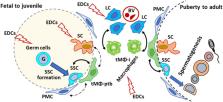
          During the past decades, several endocrine disrupting chemicals (EDCs) have been confirmed to affect male reproductive function and fertility in animal studies. EDCs are suspected to exert similar effects in humans, based on strong associations between levels of antiandrogenic EDCs in pregnant women and adverse reproductive effects in infants. Testicular macrophages (tMΦ) play a vital role in modulating immunological privilege and maintaining normal testicular homeostasis as well as fetal development. Although tMΦ were not historically studied in the context of endocrine disruption, they have emerged as potential targets to consider due to their critical role in regulating cells such as spermatogonial stem cells (SSCs) and Leydig cells. Few studies have examined the impact of EDCs on the ability of testicular cells to communicate and regulate each other’s functions. In this review, we recapitulate what is known about tMΦ functions and interactions with other cell types in the testis that support spermatogenesis and steroidogenesis. We also surveyed the literature for reports on the effects of the EDCs genistein and DEHP on tMΦ, SSCs, Sertoli and Leydig cells. Our goal is to explore the possibility that EDC disruption of tMΦ interactions with other cell types may play a role in their adverse effects on testicular developmental programming and functions. This approach will highlight gaps of knowledge, which, once resolved, should improve the risk assessment of EDC exposure and the development of safeguards to protect male reproductive functions.

          Related collections

          Most cited references155

          • Record: found
          • Abstract: found
          • Article: not found

          Exploring the full spectrum of macrophage activation.

          Macrophages display remarkable plasticity and can change their physiology in response to environmental cues. These changes can give rise to different populations of cells with distinct functions. In this Review we suggest a new grouping of macrophage populations based on three different homeostatic activities - host defence, wound healing and immune regulation. We propose that similarly to primary colours, these three basic macrophage populations can blend into various other 'shades' of activation. We characterize each population and provide examples of macrophages from specific disease states that have the characteristics of one or more of these populations.
            Bookmark
            • Record: found
            • Abstract: found
            • Article: not found

            Macrophage plasticity and polarization: in vivo veritas.

            Diversity and plasticity are hallmarks of cells of the monocyte-macrophage lineage. In response to IFNs, Toll-like receptor engagement, or IL-4/IL-13 signaling, macrophages undergo M1 (classical) or M2 (alternative) activation, which represent extremes of a continuum in a universe of activation states. Progress has now been made in defining the signaling pathways, transcriptional networks, and epigenetic mechanisms underlying M1-M2 or M2-like polarized activation. Functional skewing of mononuclear phagocytes occurs in vivo under physiological conditions (e.g., ontogenesis and pregnancy) and in pathology (allergic and chronic inflammation, tissue repair, infection, and cancer). However, in selected preclinical and clinical conditions, coexistence of cells in different activation states and unique or mixed phenotypes have been observed, a reflection of dynamic changes and complex tissue-derived signals. The identification of mechanisms and molecules associated with macrophage plasticity and polarized activation provides a basis for macrophage-centered diagnostic and therapeutic strategies.
              Bookmark
              • Record: found
              • Abstract: found
              • Article: not found

              CCL2 recruits inflammatory monocytes to facilitate breast tumor metastasis

              Macrophages abundantly found in the tumor microenvironment enhance malignancy 1 . At metastatic sites a distinct population of metastasis associated macrophages (MAMs) promote tumor cell extravasation, seeding and persistent growth 2 . Our study has defined the origin of these macrophages by showing Gr1+ inflammatory monocytes (IMs) are preferentially recruited to pulmonary metastases but not primary mammary tumors, a process also found for human IMs in pulmonary metastases of human breast cancer cells. The recruitment of these CCR2 (receptor for chemokine CCL2) expressing IMs and subsequently MAMs and their interaction with metastasizing tumor cells is dependent on tumor and stromal synthesized CCL2 (FigS1). Inhibition of CCL2/CCR2 signaling using anti-CCL2 antibodies blocks IM recruitment and inhibits metastasis in vivo and prolongs the survival of tumor-bearing mice. Depletion of tumor cell-derived CCL2 also inhibits metastatic seeding. IMs promote tumor cell extravasation in a process that requires monocyte-derived VEGF. CCL2 expression and macrophage infiltration are correlated with poor prognosis and metastatic disease in human breast cancer (Fig S2) 3-6 . Our data provides the mechanistic link between these two clinical associations and indicates new therapeutic targets for treating metastatic breast disease.
                Bookmark

                Author and article information

                Contributors
                Journal
                Front Toxicol
                Front Toxicol
                Front. Toxicol.
                Frontiers in Toxicology
                Frontiers Media S.A.
                2673-3080
                31 August 2023
                2023
                : 5
                : 1242634
                Affiliations
                Department of Pharmacology and Pharmaceutical Sciences , Alfred E. Mann School of Pharmacy and Pharmaceutical Sciences , University of Southern California , Los Angeles, CA, United States
                Author notes

                Edited by: Marina Trevizan Guerra, Federal University of Mato Grosso do Sul, Brazil

                Reviewed by: Geraldine Delbes, Institut National de la Recherche Scientifique, Canada

                Jasim Khan, University of Alabama at Birmingham, United States

                *Correspondence: Martine Culty, culty@ 123456usc.edu
                Article
                1242634
                10.3389/ftox.2023.1242634
                10501733
                37720385
                9538efe9-5970-4f0f-82c6-8c0bc4a80db2
                Copyright © 2023 Cui and Culty.

                This is an open-access article distributed under the terms of the Creative Commons Attribution License (CC BY). The use, distribution or reproduction in other forums is permitted, provided the original author(s) and the copyright owner(s) are credited and that the original publication in this journal is cited, in accordance with accepted academic practice. No use, distribution or reproduction is permitted which does not comply with these terms.

                History
                : 19 June 2023
                : 18 August 2023
                Funding
                This work was supported by funds from the University of Southern California Alfred E. Mann School of Pharmacy and Pharmaceutical Sciences to MC; and did not receive any grant from funding agencies.
                Categories
                Toxicology
                Review
                Custom metadata
                Developmental and Reproductive Toxicology

                endocrine disrupting chemicals,testis,macrophages,germ cells,leydig cells,genistein,dehp,cell-cell interactions

                Comments

                Comment on this article