26
views
0
recommends
+1 Recommend
0 collections
    0
    shares
      • Record: found
      • Abstract: found
      • Article: found
      Is Open Access

      ALPK1 hotspot mutation as a driver of human spiradenoma and spiradenocarcinoma

      research-article

      Read this article at

      Bookmark
          There is no author summary for this article yet. Authors can add summaries to their articles on ScienceOpen to make them more accessible to a non-specialist audience.

          Abstract

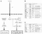
          Spiradenoma and cylindroma are distinctive skin adnexal tumors with sweat gland differentiation and potential for malignant transformation and aggressive behaviour. We present the genomic analysis of 75 samples from 57 representative patients including 15 cylindromas, 17 spiradenomas, 2 cylindroma–spiradenoma hybrid tumors, and 24 low- and high-grade spiradenocarcinoma cases, together with morphologically benign precursor regions of these cancers. We reveal somatic or germline alterations of the CYLD gene in 15/15 cylindromas and 5/17 spiradenomas, yet only 2/24 spiradenocarcinomas. Notably, we find a recurrent missense mutation in the kinase domain of the ALPK1 gene in spiradenomas and spiradenocarcinomas, which is mutually exclusive from mutation of CYLD and can activate the NF-κB pathway in reporter assays. In addition, we show that high-grade spiradenocarcinomas carry loss-of-function TP53 mutations, while cylindromas may have disruptive mutations in DNMT3A. Thus, we reveal the genomic landscape of adnexal tumors and therapeutic targets.

          Abstract

          Spiradenoma and cylindroma are skin adnexal tumors that can behave aggressively and undergo malignant transformation. Here, the authors genetically assess a cohort of these adnexal tumours, highlighting recurrent ALPK1 mutations and revealing the genomic landscape of these rare tumours.

          Related collections

          Most cited references32

          • Record: found
          • Abstract: found
          • Article: not found

          Mutations in genes encoding the cadherin receptor-ligand pair DCHS1 and FAT4 disrupt cerebral cortical development.

          The regulated proliferation and differentiation of neural stem cells before the generation and migration of neurons in the cerebral cortex are central aspects of mammalian development. Periventricular neuronal heterotopia, a specific form of mislocalization of cortical neurons, can arise from neuronal progenitors that fail to negotiate aspects of these developmental processes. Here we show that mutations in genes encoding the receptor-ligand cadherin pair DCHS1 and FAT4 lead to a recessive syndrome in humans that includes periventricular neuronal heterotopia. Reducing the expression of Dchs1 or Fat4 within mouse embryonic neuroepithelium increased progenitor cell numbers and reduced their differentiation into neurons, resulting in the heterotopic accumulation of cells below the neuronal layers in the neocortex, reminiscent of the human phenotype. These effects were countered by concurrent knockdown of Yap, a transcriptional effector of the Hippo signaling pathway. These findings implicate Dchs1 and Fat4 upstream of Yap as key regulators of mammalian neurogenesis.
            Bookmark
            • Record: found
            • Abstract: found
            • Article: found
            Is Open Access

            Function and cancer genomics of FAT family genes

            FAT1, FAT2, FAT3 and FAT4 are human homologs of Drosophila Fat, which is involved in tumor suppression and planar cell polarity (PCP). FAT1 and FAT4 undergo the first proteolytic cleavage by Furin and are predicted to undergo the second cleavage by γ-secretase to release intracellular domain (ICD). Ena/VAPS-binding to FAT1 induces actin polymerization at lamellipodia and filopodia to promote cell migration, while Scribble-binding to FAT1 induces phosphorylation and functional inhibition of YAP1 to suppress cell growth. FAT1 is repressed in oral cancer owing to homozygous deletion or epigenetic silencing and is preferentially downregulated in invasive breast cancer. On the other hand, FAT1 is upregulated in leukemia and prognosis of preB-ALL patients with FAT1 upregulation is poor. FAT4 directly interacts with MPDZ/MUPP1 to recruit membrane-associated guanylate kinase MPP5/PALS1. FAT4 is involved in the maintenance of PCP and inhibition of cell proliferation. FAT4 mRNA is repressed in breast cancer and lung cancer due to promoter hypermethylation. FAT4 gene is recurrently mutated in several types of human cancers, such as melanoma, pancreatic cancer, gastric cancer and hepatocellular carcinoma. FAT1 and FAT4 suppress tumor growth via activation of Hippo signaling, whereas FAT1 promotes tumor migration via induction of actin polymerization. FAT1 is tumor suppressive or oncogenic in a context-dependent manner, while FAT4 is tumor suppressive. Copy number aberration, translocation and point mutation of FAT1, FAT2, FAT3, FAT4, FRMD1, FRMD6, NF2, WWC1, WWC2, SAV1, STK3, STK4, MOB1A, MOB1B, LATS1, LATS2, YAP1 and WWTR1/TAZ genes should be comprehensively investigated in various types of human cancers to elucidate the mutation landscape of the FAT-Hippo signaling cascades. Because YAP1 and WWTR1 are located at the crossroads of adhesion, GPCR, RTK and stem-cell signaling network, cancer genomics of the FAT signaling cascades could be applied for diagnostics, prognostics and therapeutics in the era of personalized medicine.
              Bookmark
              • Record: found
              • Abstract: found
              • Article: not found

              Burden Testing of Rare Variants Identified through Exome Sequencing via Publicly Available Control Data

              The genetic causes of many Mendelian disorders remain undefined. Factors such as lack of large multiplex families, locus heterogeneity, and incomplete penetrance hamper these efforts for many disorders. Previous work suggests that gene-based burden testing—where the aggregate burden of rare, protein-altering variants in each gene is compared between case and control subjects—might overcome some of these limitations. The increasing availability of large-scale public sequencing databases such as Genome Aggregation Database (gnomAD) can enable burden testing using these databases as controls, obviating the need for additional control sequencing for each study. However, there exist various challenges with using public databases as controls, including lack of individual-level data, differences in ancestry, and differences in sequencing platforms and data processing. To illustrate the approach of using public data as controls, we analyzed whole-exome sequencing data from 393 individuals with idiopathic hypogonadotropic hypogonadism (IHH), a rare disorder with significant locus heterogeneity and incomplete penetrance against control subjects from gnomAD (n = 123,136). We leveraged presumably benign synonymous variants to calibrate our approach. Through iterative analyses, we systematically addressed and overcame various sources of artifact that can arise when using public control data. In particular, we introduce an approach for highly adaptable variant quality filtering that leads to well-calibrated results. Our approach “re-discovered” genes previously implicated in IHH ( FGFR1 , TACR3 , GNRHR ). Furthermore, we identified a significant burden in TYRO3 , a gene implicated in hypogonadotropic hypogonadism in mice. Finally, we developed a user-friendly software package TRAPD (Test Rare vAriants with Public Data) for performing gene-based burden testing against public databases.
                Bookmark

                Author and article information

                Contributors
                +44 (0) 1223 496862 , da1@sanger.ac.uk
                Journal
                Nat Commun
                Nat Commun
                Nature Communications
                Nature Publishing Group UK (London )
                2041-1723
                17 May 2019
                17 May 2019
                2019
                : 10
                : 2213
                Affiliations
                [1 ]ISNI 0000 0004 0606 5382, GRID grid.10306.34, Experimental Cancer Genetics, , Wellcome Trust Sanger Institute, ; Cambridge, CB10 1SA UK
                [2 ]ISNI 0000 0004 0460 0556, GRID grid.416213.3, Department of Pathology, , Maasstad Hospital, ; Maasstadweg 21, Rotterdam, 3079 DZ The Netherlands
                [3 ]GRID grid.500037.1, Dermatopathologie Friedrichshafen, ; Siemensstrasse 6/1, 88048 Friedrichshafen, Germany
                [4 ]ISNI 0000 0001 1271 4623, GRID grid.18886.3f, Dynamical Cell Systems Laboratory. Chester Beatty Laboratories, , Division of Cancer Biology. Institute of Cancer Research, ; London, SW3 6JB UK
                [5 ]ISNI 0000 0001 0721 6013, GRID grid.8954.0, Institute of Pathology, , Medical Faculty University of Ljubljana, ; Korytkova 2, Ljubljana, 1000 Slovenia
                [6 ]ISNI 0000 0004 0397 9648, GRID grid.412688.1, University Hospital Center Zagreb, ; Kispaticeva 12, 10 000 Zagreb, Croatia
                [7 ]ISNI 0000 0001 0684 291X, GRID grid.418119.4, Institut Jules Bordet, ; 121-125, boulevard de Waterloo, 1000 Brussels, Belgium
                [8 ]ISNI 0000 0001 0675 4725, GRID grid.239578.2, Department of Pathology, , Cleveland Clinic, ; Cleveland, OH 44195 USA
                [9 ]ISNI 0000 0004 1936 7697, GRID grid.22072.35, Departments of Pathology & Laboratory Medicine and Medicine and The Arnie Charbonneau Cancer Institute, Cumming School of Medicine, , University of Calgary, ; Calgary, AB T2L 2K8 Canada
                [10 ]ISNI 0000000086837370, GRID grid.214458.e, Departments of Pathology and Dermatology, , University of Michigan Medical School, ; 2800 Plymouth Road, Ann Arbor, MI 48109-5602 USA
                [11 ]ISNI 0000 0004 1936 7988, GRID grid.4305.2, Division of Pathology, Cancer Research UK Edinburgh Centre, , The University of Edinburgh, Institute of Genetics & Molecular Medicine, ; Crewe Road, Edinburgh, EH4 2XR UK
                [12 ]ISNI 0000 0001 2159 0001, GRID grid.9486.3, Laboratorio Internacional de Investigación sobre el Genoma Humano, , Universidad Nacional Autónoma de México, ; Campus Juriquilla, Blvd Juriquilla 3001, Santiago de Querétaro, 76230 Mexico
                [13 ]ISNI 000000041936754X, GRID grid.38142.3c, Department of Pathology, Brigham and Women’s Hospital, , Harvard Medical School, ; Boston, MA 02115 USA
                Author information
                http://orcid.org/0000-0002-9017-2644
                http://orcid.org/0000-0002-0413-6744
                http://orcid.org/0000-0002-6826-8770
                Article
                9979
                10.1038/s41467-019-09979-0
                6525246
                31101826
                9a652fbe-df32-4c3a-aec4-67f8e72f5920
                © The Author(s) 2019

                Open Access This article is licensed under a Creative Commons Attribution 4.0 International License, which permits use, sharing, adaptation, distribution and reproduction in any medium or format, as long as you give appropriate credit to the original author(s) and the source, provide a link to the Creative Commons license, and indicate if changes were made. The images or other third party material in this article are included in the article’s Creative Commons license, unless indicated otherwise in a credit line to the material. If material is not included in the article’s Creative Commons license and your intended use is not permitted by statutory regulation or exceeds the permitted use, you will need to obtain permission directly from the copyright holder. To view a copy of this license, visit http://creativecommons.org/licenses/by/4.0/.

                History
                : 16 November 2018
                : 8 April 2019
                Funding
                Funded by: FundRef https://doi.org/10.13039/100004440, Wellcome Trust (Wellcome);
                Funded by: FundRef https://doi.org/10.13039/501100000289, Cancer Research UK (CRUK);
                Categories
                Article
                Custom metadata
                © The Author(s) 2019

                Uncategorized
                cancer genomics,cancer genetics
                Uncategorized
                cancer genomics, cancer genetics

                Comments

                Comment on this article