50
views
0
recommends
+1 Recommend
0 collections
    0
    shares
      • Record: found
      • Abstract: found
      • Article: found
      Is Open Access

      Mesenchymal Stem/Stromal Cell-Derived Extracellular Vesicles and Their Potential as Novel Immunomodulatory Therapeutic Agents

      review-article

      Read this article at

      Bookmark
          There is no author summary for this article yet. Authors can add summaries to their articles on ScienceOpen to make them more accessible to a non-specialist audience.

          Abstract

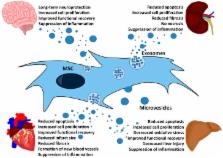
          Extracellular vesicles (EVs), such as exosomes and microvesicles, have been identified as mediators of a newly-discovered intercellular communication system. They are essential signaling mediators in various physiological and pathophysiological processes. Depending on their origin, they fulfill different functions. EVs of mesenchymal stem/stromal cells (MSCs) have been found to promote comparable therapeutic activities as MSCs themselves. In a variety of in vivo models, it has been observed that they suppress pro-inflammatory processes and reduce oxidative stress and fibrosis. By switching pro-inflammatory into tolerogenic immune responses, MSC-EVs very likely promote tissue regeneration by creating a pro-regenerative environment allowing endogenous stem and progenitor cells to successfully repair affected tissues. Accordingly, MSC-EVs provide a novel, very promising therapeutic agent, which has already been successfully applied to humans. However, the MSC-EV production process has not been standardized, yet. Indeed, a collection of different protocols has been used for the MSC-EV production, characterization and application. By focusing on kidney, heart, liver and brain injuries, we have reviewed the major outcomes of published MSC-EV in vivo studies.

          Related collections

          Most cited references35

          • Record: found
          • Abstract: found
          • Article: not found

          Administered mesenchymal stem cells protect against ischemic acute renal failure through differentiation-independent mechanisms.

          Severe acute renal failure (ARF) remains a common, largely treatment-resistant clinical problem with disturbingly high mortality rates. Therefore, we tested whether administration of multipotent mesenchymal stem cells (MSC) to anesthetized rats with ischemia-reperfusion-induced ARF (40-min bilateral renal pedicle clamping) could improve the outcome through amelioration of inflammatory, vascular, and apoptotic/necrotic manifestations of ischemic kidney injury. Accordingly, intracarotid administration of MSC (approximately 10(6)/animal) either immediately or 24 h after renal ischemia resulted in significantly improved renal function, higher proliferative and lower apoptotic indexes, as well as lower renal injury and unchanged leukocyte infiltration scores. Such renoprotection was not obtained with syngeneic fibroblasts. Using in vivo two-photon laser confocal microscopy, fluorescence-labeled MSC were detected early after injection in glomeruli, and low numbers attached at microvasculature sites. However, within 3 days of administration, none of the administered MSC had differentiated into a tubular or endothelial cell phenotype. At 24 h after injury, expression of proinflammatory cytokines IL-1beta, TNF-alpha, IFN-gamma, and inducible nitric oxide synthase was significantly reduced and that of anti-inflammatory IL-10 and bFGF, TGF-alpha, and Bcl-2 was highly upregulated in treated kidneys. We conclude that the early, highly significant renoprotection obtained with MSC is of considerable therapeutic promise for the cell-based management of clinical ARF. The beneficial effects of MSC are primarily mediated via complex paracrine actions and not by their differentiation into target cells, which, as such, appears to be a more protracted response that may become important in late-stage organ repair.
            Bookmark
            • Record: found
            • Abstract: found
            • Article: not found

            Mesenchymal stem cells suppress lymphocyte proliferation in vitro and prolong skin graft survival in vivo.

            Mesenchymal stem cells (MSCs), multipotential cells that reside within the bone marrow, can be induced to differentiate into various components of the marrow microenvironment, such as bone, adipose, and stromal tissues. The bone marrow microenvironment is vital to the development, differentiation, and regulation of the lymphohematopoietic system. We hypothesized that the activities of MSCs in the bone marrow microenvironment might also include immunomodulatory effects on lymphocytes. Baboon MSCs were tested in vitro for their ability to elicit a proliferative response from allogeneic lymphocytes, to inhibit an ongoing allogeneic response, and to inhibit a proliferative response to potent T-cell mitogens. In vivo effects were tested by intravenous administration of donor MSCs to MHC-mismatched recipient baboons prior to placement of autologous, donor, and third-party skin grafts. MSCs failed to elicit a proliferative response from allogeneic lymphocytes. MSCs added into a mixed lymphocyte reaction, either on day 0 or on day 3, or to mitogen-stimulated lymphocytes, led to a greater than 50% reduction in proliferative activity. This effect could be maximized by escalating the dose of MSCs and could be reduced with the addition of exogenous IL-2. In vivo administration of MSCs led to prolonged skin graft survival when compared to control animals: 11.3 +/- 0.3 vs 7 +/- 0. Baboon MSCs have been observed to alter lymphocyte reactivity to allogeneic target cells and tissues. These immunoregulatory features may prove useful in future applications of tissue regeneration and stem cell engineering.
              Bookmark
              • Record: found
              • Abstract: found
              • Article: not found

              Evidence supporting paracrine hypothesis for Akt-modified mesenchymal stem cell-mediated cardiac protection and functional improvement.

              We previously reported that intramyocardial injection of bone marrow-derived mesenchymal stem cells overexpressing Akt (Akt-MSCs) inhibits ventricular remodeling and restores cardiac function measured 2 wk after myocardial infarction. Here, we report that the functional improvement occurs in < 72 h. This early remarkable effect cannot be readily attributed to myocardial regeneration from the donor cells. Thus, we hypothesized that paracrine actions exerted by the cells through the release of soluble factors might be important mechanisms of tissue repair and functional improvement after injection of the Akt-MSCs. Indeed, in the current study we demonstrate that conditioned medium from hypoxic Akt-MSCs markedly inhibits hypoxia-induced apoptosis and triggers vigorous spontaneous contraction of adult rat cardiomyocytes in vitro. When injected into infarcted hearts, the Akt-MSC conditioned medium significantly limits infarct size and improves ventricular function relative to controls. Support to the paracrine hypothesis is provided by data showing that several genes, coding for factors (VEGF, FGF-2, HGF, IGF-I, and TB4) that are potential mediators of the effects exerted by the Akt-MSC conditioned medium, are significantly up-regulated in the Akt-MSCs, particularly in response to hypoxia. Taken together, our data support Akt-MSC-mediated paracrine mechanisms of myocardial protection and functional improvement.
                Bookmark

                Author and article information

                Journal
                Int J Mol Sci
                Int J Mol Sci
                ijms
                International Journal of Molecular Sciences
                MDPI
                1422-0067
                06 July 2017
                July 2017
                : 18
                : 7
                : 1450
                Affiliations
                Institute for Transfusion Medicine, University Hospital Essen, University Duisburg-Essen, Virchowstr. 179, 45147 Essen, Germany; michel.bremer@ 123456uk-essen.de (M.B.); rita.ferrertur@ 123456uk-essen.de (R.F.-T.); lena.gockeln@ 123456gmx.de (L.G.); Oumaima.Stambouli@ 123456ruhr-uni-bochum.de (O.S.); Amina.Becic@ 123456ruhr-uni-bochum.de (A.B.)
                Author notes
                [* ]Correspondence: verena.boerger@ 123456uk-essen.de (V.B.); bernd.giebel@ 123456uk-essen.de (B.G.); Tel.: +49-201-723-83038 (V.B.); +49-201-723-4204 (B.G.)
                [†]

                These authors contributed equally to this work.

                Author information
                https://orcid.org/0000-0002-8230-2218
                Article
                ijms-18-01450
                10.3390/ijms18071450
                5535941
                28684664
                9db42b96-3461-4e41-9d91-4d7b106ea557
                © 2017 by the authors.

                Licensee MDPI, Basel, Switzerland. This article is an open access article distributed under the terms and conditions of the Creative Commons Attribution (CC BY) license ( http://creativecommons.org/licenses/by/4.0/).

                History
                : 15 June 2017
                : 30 June 2017
                Categories
                Review

                Molecular biology
                mesenchymal stem/stromal cells,cell therapy,extracellular vesicles,exosomes,microvesicles,immunomodulation

                Comments

                Comment on this article