2
views
0
recommends
+1 Recommend
0 collections
    0
    shares
      • Record: found
      • Abstract: found
      • Article: found
      Is Open Access

      Establishment and genetic characterization of cell lines derived from proliferating nasal polyps and sinonasal inverted papillomas

      research-article

      Read this article at

      Bookmark
          There is no author summary for this article yet. Authors can add summaries to their articles on ScienceOpen to make them more accessible to a non-specialist audience.

          Abstract

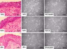
          To better understand the pathogenesis of nasal polyps (NPs) and sinonasal inverted papillomas (SIPs), we aimed to establish cell lines from fresh tissues of NPs and SIPs and characterize them. Primary cell cultures were obtained from two NP tissues (NP2 and NP3) and one SIP tissue (IP4). All the cells were polygonal in shape, expressed cytokeratin 14, and had normal diploid chromosome status. HPV58 DNA was detected in NP3. To obtain immortal primary cells, NP2 and IP4 cells were transduced with a combination of mutant CDK4, cyclinD1 and TERT. These cells were thereafter named NP2/K4DT and IP4/K4DT, respectively. HPV58-positive NP3 cells were transduced with TERT alone, the resulting cells named NP3/T. Phenotypic and genotypic identity of original tissues and derived cells was investigated. All the cell cultures with transgenes were confirmed to be derived from their parental cells and primary tumor tissues by analysis of short tandem repeats (STR) and maintained in vitro growth, genetic profiles and gene expression characteristics of the primary cells. These virtually immortalized cells, as well as the primary cells, have potential as in vitro models for studying the pathogenesis of NPs and SIPs and for preclinical study to develop new therapeutic agents.

          Related collections

          Most cited references66

          • Record: found
          • Abstract: found
          • Article: found
          Is Open Access

          Comprehensive genomic characterization of head and neck squamous cell carcinomas

          The Cancer Genome Atlas profiled 279 head and neck squamous cell carcinomas (HNSCCs) to provide a comprehensive landscape of somatic genomic alterations. We find that human papillomavirus-associated (HPV) tumors are dominated by helicase domain mutations of the oncogene PIK3CA, novel alterations involving loss of TRAF3, and amplification of the cell cycle gene E2F1. Smoking-related HNSCCs demonstrate near universal loss of TP53 mutations and CDKN2A with frequent copy number alterations including a novel amplification of 11q22. A subgroup of oral cavity tumors with favorable clinical outcomes displayed infrequent CNAs in conjunction with activating mutations of HRAS or PIK3CA, coupled with inactivating mutations of CASP8, NOTCH1 and wild-type TP53. Other distinct subgroups harbored novel loss of function alterations of the chromatin modifier NSD1, Wnt pathway genes AJUBA and FAT1, and activation of oxidative stress factor NFE2L2, mainly in laryngeal tumors. Therapeutic candidate alterations were identified in the majority of HNSCC's.
            Bookmark
            • Record: found
            • Abstract: found
            • Article: not found

            The role of senescent cells in ageing.

            Cellular senescence has historically been viewed as an irreversible cell-cycle arrest mechanism that acts to protect against cancer, but recent discoveries have extended its known role to complex biological processes such as development, tissue repair, ageing and age-related disorders. New insights indicate that, unlike a static endpoint, senescence represents a series of progressive and phenotypically diverse cellular states acquired after the initial growth arrest. A deeper understanding of the molecular mechanisms underlying the multi-step progression of senescence and the development and function of acute versus chronic senescent cells may lead to new therapeutic strategies for age-related pathologies and extend healthy lifespan.
              Bookmark
              • Record: found
              • Abstract: found
              • Article: not found

              Serial cultivation of strains of human epidermal keratinocytes: the formation of keratinizing colonies from single cells.

              Human diploid epidermis epidermal cells have been successfully grown in serial culture. To initiate colony formation, they require the presence of fibroblasts, but proliferation of fibroblasts must be controlled so that the epidermal cell population is not overgrown. Both conditions can be achieved by the use of lethally irradiated 3T3 cells at the correct density. When trypsinized human skin cells are plated together with the 3T3 cells, the growth of the human fibroblasts is largely suppressed, but epidermal cells grow from single cells into colonies. Each colony consists of keratinocytes ultimately forming a stratified squamous epithelium in which the dividing cells are confined to the lowest layer(s). Hydrocortisone is added to the medium, since in secondary and subsequent subcultures it makes the colony morphology more oderly and distinctive, and maintains proliferation at a slightly greater rate. Under these culture conditions, it is possible to isolate keratinocyte clones free of viable fibroblasts. Like human diploid fibroblasts, human diploid keratinocytes appear to have a finite culture lifetime. For 7 strains studied, the culture lifetime ranged from 20-50 cell generations. The plating efficiency of the epidermal cells taken directly from skin was usually 0.1-1.0%. On subsequent transfer of the cultures initiated from newborns, the plating efficiency rose to 10% or higher, but was most often in the range of 1-5% and dropped sharply toward the end of their culture life. The plating efficiency and culture lifetime were lower for keratinocytes of older persons.
                Bookmark

                Author and article information

                Contributors
                tkiyono@east.ncc.go.jp
                chapie@kku.ac.th
                Journal
                Sci Rep
                Sci Rep
                Scientific Reports
                Nature Publishing Group UK (London )
                2045-2322
                24 August 2021
                24 August 2021
                2021
                : 11
                : 17100
                Affiliations
                [1 ]GRID grid.9786.0, ISNI 0000 0004 0470 0856, Department of Microbiology, Faculty of Medicine, , Khon Kaen University, ; Khon Kaen, Thailand
                [2 ]GRID grid.9786.0, ISNI 0000 0004 0470 0856, HPV & EBV and Carcinogenesis Research Group, , Khon Kaen University, ; Khon Kaen, Thailand
                [3 ]GRID grid.272242.3, ISNI 0000 0001 2168 5385, Project for Prevention of HPV-Related Cancer, Exploratory Oncology Research and Clinical Trial Center, , National Cancer Center, ; 6-5-1 Kashiwanoha, Kashiwa, Chiba 277-8577 Japan
                [4 ]GRID grid.9786.0, ISNI 0000 0004 0470 0856, Department of Otorhinolaryngology, Faculty of Medicine, , Khon Kaen University, ; Khon Kaen, Thailand
                [5 ]GRID grid.9786.0, ISNI 0000 0004 0470 0856, Department of Pathology, Faculty of Medicine, , Khon Kaen University, ; Khon Kaen, Thailand
                [6 ]GRID grid.9786.0, ISNI 0000 0004 0470 0856, Department of Forensic Medicine, Faculty of Medicine, , Khon Kaen University, ; Khon Kaen, Thailand
                Article
                96444
                10.1038/s41598-021-96444-y
                8384845
                34429452
                9f26f787-4c38-4b71-beeb-8f7bfa85f2cd
                © The Author(s) 2021

                Open Access This article is licensed under a Creative Commons Attribution 4.0 International License, which permits use, sharing, adaptation, distribution and reproduction in any medium or format, as long as you give appropriate credit to the original author(s) and the source, provide a link to the Creative Commons licence, and indicate if changes were made. The images or other third party material in this article are included in the article's Creative Commons licence, unless indicated otherwise in a credit line to the material. If material is not included in the article's Creative Commons licence and your intended use is not permitted by statutory regulation or exceeds the permitted use, you will need to obtain permission directly from the copyright holder. To view a copy of this licence, visit http://creativecommons.org/licenses/by/4.0/.

                History
                : 6 January 2021
                : 10 August 2021
                Funding
                Funded by: The Thailand Research Fund and Khon Kaen University through the Royal Golden Jubilee Ph.D. Program
                Award ID: PHD/0081/2558
                Award Recipient :
                Funded by: FundRef http://dx.doi.org/10.13039/501100004071, Khon Kaen University;
                Award ID: B05F630053
                Award Recipient :
                Funded by: Japan Agency for Medical Research
                Award ID: 20jk0210009
                Award Recipient :
                Funded by: Research and Graduate Studies, Khon Kaen University
                Award ID: RP64-4/004
                Award Recipient :
                Categories
                Article
                Custom metadata
                © The Author(s) 2021

                Uncategorized
                cancer models,head and neck cancer
                Uncategorized
                cancer models, head and neck cancer

                Comments

                Comment on this article