19
views
0
recommends
+1 Recommend
0 collections
    0
    shares
      • Record: found
      • Abstract: found
      • Article: found
      Is Open Access

      Effect of mild hypothermia on lung injury after cardiac arrest in swine based on lung ultrasound

      research-article

      Read this article at

      Bookmark
          There is no author summary for this article yet. Authors can add summaries to their articles on ScienceOpen to make them more accessible to a non-specialist audience.

          Abstract

          Background

          Lung injury is common in post-cardiac arrest syndrome, and is associated with increased morbidity and mortality. The aim of this study was to evaluate the effect of mild hypothermia on lung injury after cardiac arrest in swine based on lung ultrasound.

          Methods

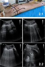
          Twenty-three male domestic swine weighing 36 ± 2 kg were randomly assigned to three groups: therapeutic hypothermia (TH, n = 9), normothermia (NT, n = 9), and sham control (control, n = 5) groups. Sham animals only underwent surgical preparation. The animal model was established with 8 min of ventricular fibrillation followed by 5 min of cardiopulmonary resuscitation. Therapeutic hypothermia was induced and maintained until 24 h post-resuscitation in the TH group by surface blanket cooling, followed by rewarming at a rate of 1 °C/h for 5 h. The extravascular lung water index (ELWI), pulmonary vascular permeability index (PVPI), PO 2/FiO 2, and lung ultrasound score (LUS) were measured at baseline and at 1, 3, 6, 12, 24, and 30 h after resuscitation. After euthanizing the swine, their lung tissues were quickly obtained to evaluate inflammation.

          Results

          After resuscitation, ELWI and PVPI in the NT group were higher, and PO 2/FiO 2 was lower, than in the sham group. However, those measures were significantly better in the TH group than the NT group. The LUS was higher in the NT group than in the sham group at 1, 3, 6, 12, 24, and 30 h after resuscitation. The LUS was significantly better in the TH group compared to the NT group. The lung tissue biopsy revealed that lung injury was more severe in the NT group than in the TH group. Increases in LUS were highly correlated with increases in ELWI (r = 0.613; p < 0.001) and PVPI (r = 0.683; p < 0.001), and decreases in PO 2/FiO 2 (r = − 0.468; p < 0.001).

          Conclusions

          Mild hypothermia protected against post-resuscitation lung injury in a swine model of cardiac arrest. Lung ultrasound was useful to dynamically evaluate the role of TH in lung protection.

          Related collections

          Most cited references28

          • Record: found
          • Abstract: found
          • Article: not found

          The comet-tail artifact. An ultrasound sign of alveolar-interstitial syndrome.

          Can ultrasound be of any help in the diagnosis of alveolar-interstitial syndrome? In a prospective study, we examined 250 consecutive patients in a medical intensive care unit: 121 patients with radiologic alveolar-interstitial syndrome (disseminated to the whole lung, n = 92; localized, n = 29) and 129 patients without radiologic evidence of alveolar-interstitial syndrome. The antero-lateral chest wall was examined using ultrasound. The ultrasonic feature of multiple comet-tail artifacts fanning out from the lung surface was investigated. This pattern was present all over the lung surface in 86 of 92 patients with diffuse alveolar-interstitial syndrome (sensitivity of 93.4%). It was absent or confined to the last lateral intercostal space in 120 of 129 patients with normal chest X-ray (specificity of 93.0%). Tomodensitometric correlations showed that the thickened sub-pleural interlobular septa, as well as ground-glass areas, two lesions present in acute pulmonary edema, were associated with the presence of the comet-tail artifact. In conclusion, presence of the comet-tail artifact allowed diagnosis of alveolar-interstitial syndrome.
            Bookmark
            • Record: found
            • Abstract: found
            • Article: not found

            Successful cardiopulmonary resuscitation after cardiac arrest as a "sepsis-like" syndrome.

            We investigated the immunoinflammatory profile of patients successfully resuscitated after cardiac arrest, representing a model of whole-body ischemia/reperfusion syndrome. Plasma cytokine, endotoxin, and ex vivo cytokine production in whole-blood assays was assessed in 61, 35, and 11 patients, respectively. On admission, high levels of plasma interleukin (IL)-6, IL-8, IL-10, and soluble tumor necrosis factor (TNF) receptor type II could discriminate between survivors and nonsurvivors. Among nonsurvivors, the initial need for a vasopressor agent was associated with higher levels of IL-1 receptor antagonist, IL-10, and IL-6 on day 1. Plasma endotoxin was detected in 46% of the analyzed patients within the 2 first days. Endotoxin-induced TNF and IL-6 productions were dramatically impaired in these patients compared with healthy control subjects, whereas an unaltered production was observed with heat-killed Staphylococcus aureus. In contrast, IL-1 receptor antagonist productions were enhanced in these patients compared with healthy control subjects. The productions of T-cell-derived IL-10 and interferon-gamma were also impaired in these patients. Finally, using in vitro plasma exchange between healthy control subjects and patients, we demonstrated that the endotoxin-dependent hyporeactivity was an intrinsic property of patients' leukocytes and that an immunosuppressive activity was also present in their plasma. Altogether, the high levels of circulating cytokines, the presence of endotoxin in plasma, and the dysregulated production of cytokines found in these patients recall the immunological profile found in patients with sepsis.
              Bookmark
              • Record: found
              • Abstract: found
              • Article: not found

              Lung Ultrasound for Critically Ill Patients

              Point-of-care ultrasound is increasingly used at the bedside to integrate the clinical assessment of the critically ill; in particular, lung ultrasound has greatly developed in the last decade. This review describes basic lung ultrasound signs and focuses on their applications in critical care. Lung semiotics are composed of artifacts (derived by air/tissue interface) and real images (i.e., effusions and consolidations), both providing significant information to identify the main acute respiratory disorders. Lung ultrasound signs, either alone or combined with other point-of-care ultrasound techniques, are helpful in the diagnostic approach to patients with acute respiratory failure, circulatory shock, or cardiac arrest. Moreover, a semiquantification of lung aeration can be performed at the bedside and used in mechanically ventilated patients to guide positive end-expiratory pressure setting, assess the efficacy of treatments, monitor the evolution of the respiratory disorder, and help the weaning process. Finally, lung ultrasound can be used for early detection and management of respiratory complications under mechanical ventilation, such as pneumothorax, ventilator-associated pneumonia, atelectasis, and pleural effusions. Lung ultrasound is a useful diagnostic and monitoring tool that might in the near future become part of the basic knowledge of physicians caring for the critically ill patient.
                Bookmark

                Author and article information

                Contributors
                21518193@zju.edu.cn
                adams_ok@126.com
                jxh12199@163.com
                cqjgjc@163.com
                lizilong55@vip.sina.com
                z2jzk@zju.edu.cn
                Journal
                BMC Pulm Med
                BMC Pulm Med
                BMC Pulmonary Medicine
                BioMed Central (London )
                1471-2466
                5 November 2019
                5 November 2019
                2019
                : 19
                : 198
                Affiliations
                [1 ]ISNI 0000 0004 1759 700X, GRID grid.13402.34, Department of Emergency Medicine, Second Affiliated Hospital, Zhejiang University School of Medicine, , Institute of Emergency Medicine, Zhejiang University, ; Hangzhou, China
                [2 ]Research fellow, from Department of Emergency Medicine, Yuyao People’s Hospital, Ningbo, China
                [3 ]Research fellow, from Department of Emergency Medicine, Wenling People’s Hospital, Taizhou, China
                [4 ]Research fellow, from Department of Emergency Medicine, Ninghai People’s Hospital, Ningbo, China
                Author information
                http://orcid.org/0000-0001-7919-8213
                Article
                958
                10.1186/s12890-019-0958-8
                6833209
                31690318
                a085389d-be32-40a7-b0af-79e3ebcf9dca
                © The Author(s). 2019

                Open AccessThis article is distributed under the terms of the Creative Commons Attribution 4.0 International License ( http://creativecommons.org/licenses/by/4.0/), which permits unrestricted use, distribution, and reproduction in any medium, provided you give appropriate credit to the original author(s) and the source, provide a link to the Creative Commons license, and indicate if changes were made. The Creative Commons Public Domain Dedication waiver ( http://creativecommons.org/publicdomain/zero/1.0/) applies to the data made available in this article, unless otherwise stated.

                History
                : 3 November 2018
                : 14 October 2019
                Funding
                Funded by: 2015 Welfare Scientific Research Project from the Chinese Ministry of Health
                Award ID: 2015SQ00050
                Award Recipient :
                Funded by: Key joint research project of Chinese Ministry of Health & Zhejiang Province
                Award ID: 2018271879
                Award Recipient :
                Funded by: Welfare scientific research project of Zhejiang Province
                Award ID: LGF18H150003
                Award Recipient :
                Funded by: FundRef http://dx.doi.org/10.13039/501100001809, National Natural Science Foundation of China;
                Award ID: 81571916
                Award ID: 81372079
                Award Recipient :
                Categories
                Research Article
                Custom metadata
                © The Author(s) 2019

                Respiratory medicine
                cardiac arrest,lung injury,lung ultrasound,post-cardiac arrest syndrome,therapeutic hypothermia

                Comments

                Comment on this article